. 2011 Sep 30;286(39):34298-310.
doi: 10.1074/jbc.M111.254938. Epub 2011 Aug 2.

Pericellular versican regulates the fibroblast-myofibroblast transition: a role for ADAMTS5 protease-mediated proteolysis

Affiliations

Pericellular versican regulates the fibroblast-myofibroblast transition: a role for ADAMTS5 protease-mediated proteolysis

Noriko Hattori et al. J Biol Chem. .

Abstract

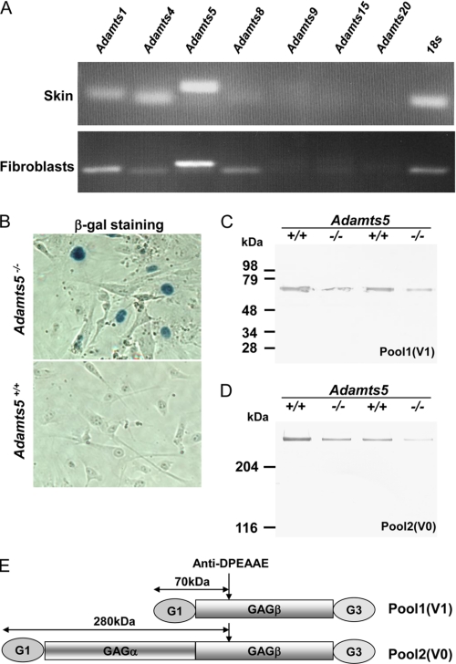
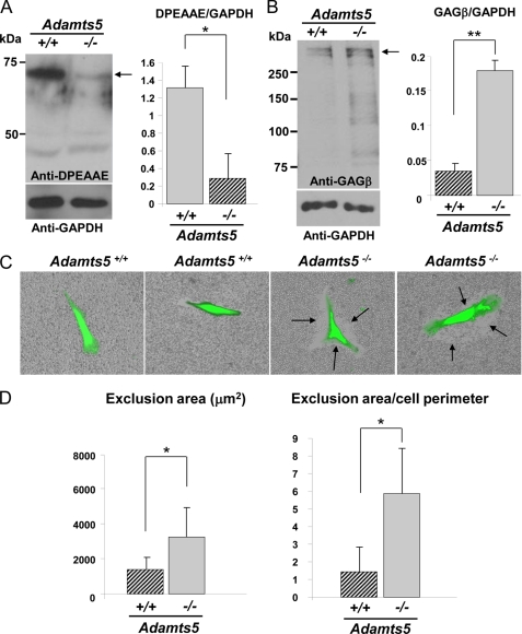
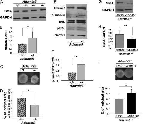
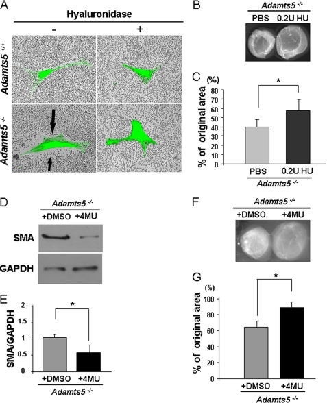
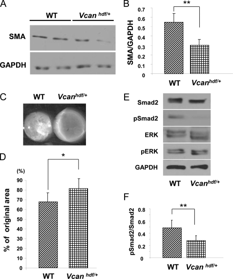
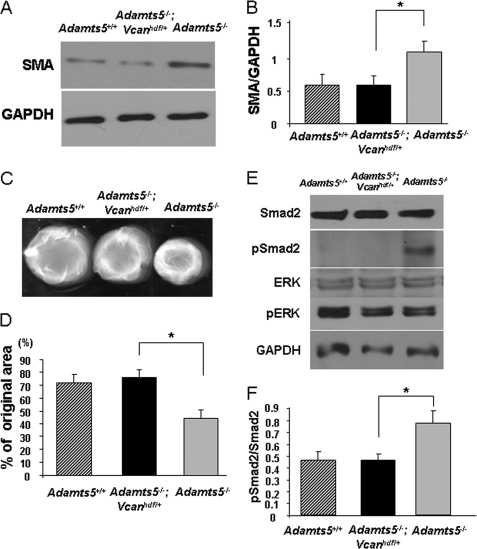
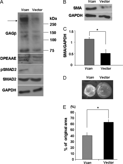
The cell and its glycosaminoglycan-rich pericellular matrix (PCM) comprise a functional unit. Because modification of PCM influences cell behavior, we investigated molecular mechanisms that regulate PCM volume and composition. In fibroblasts and other cells, aggregates of hyaluronan and versican are found in the PCM. Dermal fibroblasts from Adamts5(-/-) mice, which lack a versican-degrading protease, ADAMTS5, had reduced versican proteolysis, increased PCM, altered cell shape, enhanced α-smooth muscle actin (SMA) expression and increased contractility within three-dimensional collagen gels. The myofibroblast-like phenotype was associated with activation of TGFβ signaling. We tested the hypothesis that fibroblast-myofibroblast transition in Adamts5(-/-) cells resulted from versican accumulation in PCM. First, we noted that versican overexpression in human dermal fibroblasts led to increased SMA expression, enhanced contractility, and increased Smad2 phosphorylation. In contrast, dermal fibroblasts from Vcan haploinsufficient (Vcan(hdf/+)) mice had reduced contractility relative to wild type fibroblasts. Using a genetic approach to directly test if myofibroblast transition in Adamts5(-/-) cells resulted from increased PCM versican content, we generated Adamts5(-/-);Vcan(hdf/+) mice and isolated their dermal fibroblasts for comparison with dermal fibroblasts from Adamts5(-/-) mice. In Adamts5(-/-) fibroblasts, Vcan haploinsufficiency or exogenous ADAMTS5 restored normal fibroblast contractility. These findings demonstrate that altering PCM versican content through proteolytic activity of ADAMTS5 profoundly influenced the dermal fibroblast phenotype and may regulate a phenotypic continuum between the fibroblast and its alter ego, the myofibroblast. We propose that a physiological function of ADAMTS5 in dermal fibroblasts is to maintain optimal versican content and PCM volume by continually trimming versican in hyaluronan-versican aggregates.


Figures

FIGURE 1.
FIGURE 1.
Mouse dermal fibroblasts express Adamts5 and two versican isoforms. A, RT-PCR analyses of 3-week-old mouse skin RNA (upper panel) and of cultured WT dermal fibroblast RNA (lower panel) show expression of Adamts5 in RNA samples from both sources. Adamts1, Adamts4, and Adamts8 mRNA were also detectable in cultured fibroblasts. 18 S RNA PCR was used as a control for PCR amplification. B, β-galactosidase histochemistry of cultured WT and Adamts5−/− dermal fibroblasts shows nuclear β-galactosidase staining of Adamts5−/− dermal cells (blue nuclei) but not WT dermal cells. C and D, anti-DPEAAE Western blot of proteoglycans isolated from the skin of mice of the indicated phenotypes in pairwise analysis of Adamts5−/− mice and WT littermates is shown. The distinction between pool 1 and pool 2 is explained under “Experimental Procedures.” The DPEAAE-reactive cleaved versican arising from the V1 isoform (pool 1) and the V0 isoform (pool 2) is reduced in Adamts5−/− mice when compared with WT littermates. E, graphic representation of the molecular species identified in pool 1 and pool 2 is shown.
FIGURE 2.
FIGURE 2.
Reduced versican processing and accumulation of the PCM around cultured Adamts5−/−dermal fibroblasts. A, left-hand panel, a Western blot using anti-DPEAAE identified a 70-kDa reactive species (arrow) in WT fibroblast extracts, but this was greatly reduced in null fibroblasts. The 40-kDa species is nonspecific. The lower immunoblot shows a Western blot with GAPDH as a control (representative of n = 3). Right-hand panel, quantification of anti-DPEAAE Western blots after normalization to GAPDH shows a statistically significant difference between Adamts5−/− and WT fibroblasts (*, p < 0.01). B, left-hand panel, Western blot with an antibody to versican (anti-GAGβ) identified a higher amount of intact versican (arrow) in null fibroblasts compared with wild type. The lower panel shows a Western blot with GAPDH as a control. Versican migrates as a ∼350-kDa species when deglycosylated with chondroitinase ABC (representative of n = 4). The observed ∼350 kDa doublet may be a mixture of intact versican V1 and versican V1 lacking the N-terminal 70-kDa species. Right-hand panel, quantification of anti-GAGβ Western blots after normalization to GAPDH shows a statistically significant difference between Adamts5−/− and WT fibroblasts (**, p < 0.05). C, exclusion of RBCs around calcein-labeled fibroblasts shows accumulation of PCM around fibroblasts (representative of n = 25). The exclusion zone is indicated by arrows. D, quantification of the area of RBC exclusion (left-hand panel) and of the ratio of the exclusion area to cell perimeter (right-hand panel) shows increased PCM around Adamts5−/− dermal fibroblasts (n = 25). Error bars indicate S.D.
FIGURE 3.
FIGURE 3.
Adamts5−/− dermal fibroblasts show features of myofibroblasts. A, Western blot for SMA shows increased levels in Adamts5−/− dermal fibroblasts (−/−) compared with WT (+/+) (representative of three biological replicates and five technical replicates). B, results of densitometric analysis of SMA Western blots show a statistically significant increase in Adamts5−/− dermal fibroblasts (*, p < 0.01). C, a collagen gel contraction assay (representative example) shows the greater contractility of Adamts5−/−dermal fibroblasts. D, quantification of gel contraction assay shows a statistically significant difference (*, p < 0.01) in contractility between Adamts5−/− and WT dermal fibroblasts (n = 3 biological replicates, 4 technical replicates, and triplicate wells per experiment). E, Western blot analyses with the indicated antibodies show increased pSmad levels but no change in pERK in Adamts5−/− dermal fibroblasts (representative of n = 3). F, quantification by densitometric analysis of pSmad2/3 to total Smad2/3 shows a statistically significant difference between Adamts5−/− and WT dermal fibroblasts (*, p < 0.01) (n = 3 biological replicates). Error bars indicate S.D. G, Adamts5−/− cells were treated with SB431542, which led to decreased expression of SMA (representative of n = 3). H, quantification of SMA showed statistically significant reduction by 4MU-treated cells (**, p < 0.05). I, the collagen gel contraction assay was done using Adamts5−/− cells treated with SB431542 or with DMSO (vehicle for SB431542 delivery) as a control. J, a statistically significant reduction of contractility was observed in SB431542-treated Adamts5−/− cells (*, p < 0.01, n = 3). Error bars indicate S.D.
FIGURE 4.
FIGURE 4.
The increased PCM and altered phenotype of Adamts5−/− dermal fibroblasts can be modulated by HA content. A, treatment of Adamts5−/− or WT dermal fibroblasts with Streptomyces hyaluronidase (+) leads to loss of the PCM seen in untreated Adamts5−/− cells (−, arrows) as shown by the RBC assay (representative of n = 3). B, Adamts5−/− dermal fibroblasts embedded in collagen gels and cultured in the presence of 0.2 units (U)/ml hyaluronidase showed reduced contractility compared with untreated Adamts5−/− dermal fibroblasts. C, quantification of collagen gel contraction shows statistically significant reduction in contractility in Adamts5−/− cells treated with hyaluronidase (*, p < 0.01) (2 independent experiments with four replicates each). D, Adamts5−/− cells were treated with 4MU, which led to decreased expression of SMA (representative of n = 3). E, quantification of SMA showed statistically significant reduction in contractility by 4MU-treated cells (*, p < 0.01). F, the collagen gel contraction assay was done using Adamts5−/− cells treated with 4MU or with DMSO (vehicle for 4MU delivery) as a control. G, a statistically significant reduction of contractility was observed in 4MU-treated Adamts5−/− cells (*, p < 0.01, n = 3). Error bars indicate S.D.
FIGURE 5.
FIGURE 5.
Vcanhdf/+ dermal fibroblasts have reduced SMA, reduced contractility, and decreased pSMAD2 activation in vitro. A, a Western blot shows reduced SMA levels in Vcanhdf/+ dermal fibroblasts (representative of three biological replicates and five technical replicates; see also supplemental Fig. 3). B, quantification of SMA levels shows a statistically significant reduction of SMA in Vcanhdf/+ dermal fibroblasts (, **p < 0.05). C, a representative collagen gel contraction assay shows reduced ability of Vcanhdf/+ dermal fibroblasts to contract a collagen gel compared with WT fibroblasts. D, quantification of collagen gel contraction shows a statistically significant reduction in contractility by Vcanhdf/+ dermal fibroblasts compared with WT fibroblasts (*, p < 0.01, n = 3 biological replicates with triplicate wells per experiment). E, Western blot analysis shows decreased pSmad2 levels in Vcanhdf/+ dermal fibroblasts but no effect on pERK levels. F, quantification of the ratio of pSmad2 to total Smad2 (obtained by densitometry of Western blots) shows a statistically significant decrease of Smad2 phosphorylation in Vcanhdf/+ dermal fibroblasts (*, p < 0.01, n = 3 biological replicates).
FIGURE 6.
FIGURE 6.
Vcan haploinsufficiency abrogates the myofibroblast phenotype of Adamts5−/− dermal fibroblasts. A, a representative Western blot analysis for SMA shows that the enhanced levels seen in Adamts5−/− cells are restored to those of WT fibroblasts by Vcan haploinsufficiency. B, quantification of SMA by densitometry of Western blots shows restoration of the increased levels in Adamts5−/− cells to WT levels by Vcan haploinsufficiency (n = 3). C, representative collagen gel contraction assay shows that the increased contractility of Adamts5−/− cells is reduced by Vcan haploinsufficiency, to be similar to that of WT cells. D, quantification of collagen gel contraction illustrates the restoration of contractility to the level of WT fibroblasts in Adamts5−/−;Vcanhdf/+ cells. E, Western blot analysis shows that increased pSmad2 in Adamts5−/− cells is reduced by Vcan haploinsufficiency. F, quantification of the ratio of pSmad2 to total Smad2 (obtained by densitometry of Western blots) shows a statistically significant decrease in Smad2 phosphorylation in Adamts5−/−;Vcanhdf/+ dermal fibroblasts compared with Adamts5−/− fibroblasts. Error bars indicate S.D.
FIGURE 7.
FIGURE 7.
Versican overexpression induces transition of normal human dermal fibroblasts to a myofibroblast phenotype. A, Western blotting of cell monolayers with an antibody to versican (anti-GAGβ) shows that increased versican V1 isoform expression (arrow) was achieved by transient transfection. Versican migrates as an ∼350-kDa species when deglycosylated with chondroitinase ABC (arrow, upper panel) Coincident with this, there were increased levels of DPEAAE and pSMAD2 immunoreactivity (representative of n = 2). B, Vcan-overexpressing cells had increased levels of SMA (center panel) (representative of n = 3). C, quantification of SMA by densitometry of Western blots shows expression of significantly higher levels in normal human dermal fibroblasts by Vcan overexpression (*, p < 0.01, n = 3). D, collagen gel contraction assay showed that Vcan-overexpressing cells can contract a collagen gel to a greater extent. E, quantification of collagen gel contraction assay shows that V1 versican-overexpressing cells are contractile to a greater extent than control vector-transfected cells (*, p < 0.01, n = 3 independent transfections with three gels per experiment). Error bars indicate S.D.

Similar articles

Cited by

References

    1. Clarris B. J., Fraser J. R. (1967) Nature 214, 1159 - PubMed
    1. Clarris B. J., Fraser J. R. (1968) Exp. Cell Res. 49, 181–193 - PubMed
    1. Bennett H. S. (1963) J. Histochem. Cytochem. 11, 14–23
    1. Camaioni A., Salustri A., Yanagishita M., Hascall V. C. (1996) Arch. Biochem. Biophys. 325, 190–198 - PubMed
    1. Evanko S. P., Tammi M. I., Tammi R. H., Wight T. N. (2007) Adv. Drug Deliv. Rev. 59, 1351–1365 - PMC - PubMed

Publication types

MeSH terms

LinkOut - more resources