. 2011 Sep 23;286(38):33178-89.
doi: 10.1074/jbc.M111.253369. Epub 2011 Aug 2.

Characterization of the dimerization interface of membrane type 4 (MT4)-matrix metalloproteinase

Affiliations

Characterization of the dimerization interface of membrane type 4 (MT4)-matrix metalloproteinase

Anjum Sohail et al. J Biol Chem. .

Abstract

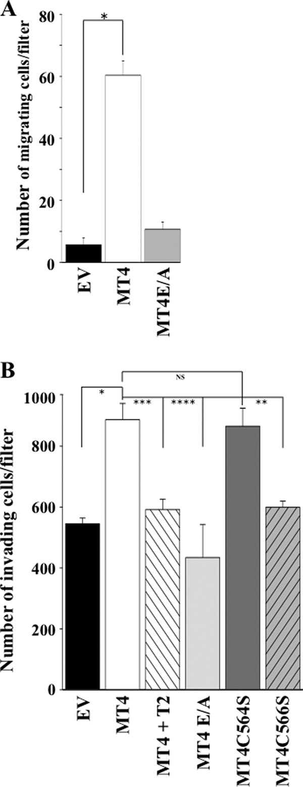
MT4-MMP (MMP17) belongs to a unique subset of membrane type-matrix metalloproteinases that are anchored to the cell surface via a glycosylphosphatidylinositol moiety. However, little is known about its biochemical properties. Here, we report that MT4-MMP is displayed on the cell surface as a mixed population of monomeric, dimeric, and oligomeric forms. Sucrose gradient fractionation demonstrated that these forms of MT4-MMP are all present in lipid rafts. Mutational and computational analyses revealed that Cys(564), which is present within the stem region, mediates MT4-MMP homodimerization by forming a disulfide bond. Substitution of Cys(564) results in a more rapid MT4-MMP turnover, when compared with the wild-type enzyme, consistent with a role for dimerization in protein stability. Expression of MT4-MMP in Madin-Darby canine kidney cells enhanced cell migration and invasion of Matrigel, a process that requires catalytic activity. However, a serine substitution at Cys(564) did not reduce MT4-MMP-stimulated cell invasion of Matrigel suggesting that homodimerization is not required for this process. Deglycosylation studies showed that MT4-MMP is modified by N-glycosylation. Moreover, inhibition of N-glycosylation by tunicamycin diminished the extent of MT4-MMP dimerization suggesting that N-glycans may confer stability to the dimeric form. Taken together, the data presented here provide a new insight into the characteristics of MT4-MMP and highlight the common and distinct properties of the glycosylphosphatidylinositol-anchored membrane type-matrix metalloproteinases.


Figures

FIGURE 1.
FIGURE 1.
Profile of MT4-MMP species. A, mouse brain (lane 1) and uterus (lane 2) tissues were homogenized in cold lysis buffer as described under “Experimental Procedures.” Homogenates (80 μg/lane) were resolved by 7.5% (−β-ME, upper panel) or 10% (+β-ME, lower panel) SDS-PAGE followed by transfer to a nitrocellulose membrane. Membranes were probed with r-mAb EP1270Y directed against the catalytic domain of MT4-MMP. B, MDCK-MT4 (lanes 2 and 4) and MDCK-EV (lanes 1 and 3) cells were surface-biotinylated as described under “Experimental Procedures.” The cells were lysed in 200 μl of cold lysis buffer, and protein concentrations were determined. One-third of the total lysate from each treatment was kept in the cold (total lysate); whereas the rest of each fraction was subjected to the pulldown assay of NeutrAvidin beads. The bound proteins were eluted with 2× Laemmli SDS sample buffer and divided in 2 aliquots; one received β-ME, and the other did not. The total lysates were also divided in two fractions; one was supplemented with β-ME and the other was not. The samples were resolved by 7.5% SDS-PAGE for reducing conditions (upper panel) and 10% SDS-PAGE for nonreducing conditions (lower panel) and then processed for immunoblot analysis using antibody EP1270Y. C, crude plasma membranes were prepared from MDCK-EV and MDCK-MT4 cells as described under “Experimental Procedures.” Twenty μg of each plasma membrane fraction were resolved by 7.5% (−β-ME, upper panel) or 10% (+β-ME, lower panel) SDS-PAGE followed by immunoblot analysis with antibody EP1270Y. D, MDCK-EV and MDCK-MT4 cells were treated with or without 0.3 IU/well of PI-PLC. After 30 min, the PI-PLC-released fraction was collected, clarified, and concentrated as described under “Experimental Procedures.” An aliquot of each concentrated supernatant was mixed with Laemmli SDS-sample buffer with or without β-ME, boiled, and resolved by 7.5% (−β-ME, upper panel) or 10% (+β-ME, lower panel) SDS-PAGE followed by immunoblot analysis with r-mAb EP1270Y. MT4O, MT4-MMP oligomeric form; MT4D, MT4-MMP dimeric form; MT4M, MT4-MMP monomeric form; pro-MT4, precursor/latent MT4-MMP.
FIGURE 2.
FIGURE 2.
MT4-MMP is targeted to lipid rafts. MDCK-MT4 cells were homogenized in cold lysis buffer containing 1% Triton X-100 and protease inhibitors as described under “Experimental Procedures” and subjected to step sucrose gradient. After centrifugation at 100,000 × g for 24 h at 4 °C, nine fractions, 1 ml of each, were collected from the top of the gradient, and an equal volume of each fraction was mixed with 4× Laemmli SDS-sample buffer with or without β-ME. The samples were resolved by 7.5% (−β-ME, upper panel) or 10% (+β-ME, lower panel) SDS-PAGE followed by immunoblot analysis with r-mAb EP1270Y. The blot with reduced protein samples was re-probed with a polyclonal antibody to human caveolin. MT4O, MT4-MMP oligomeric form; MT4D, MT4-MMP dimeric form; MT4M, MT4-MMP monomeric form.
FIGURE 3.
FIGURE 3.
MT4-MMP is N-glycosylated. A–C, MDCK-MT4 cells were treated with either BGN (A) or tunicamycin (B and C), as described under “Experimental Procedures.” A, lysates of MDCK-MT4 cells treated with either DMSO (lane 1) or 2 mm BGN (lane 2). Ctrl, control. B, lysates of MDCK-MT4 cells treated with either DMSO (lane 1) or two concentrations of tunicamycin (Tunic.) (lane 2, 5 μg/ml; lane 3, 1 μg/ml). C, lysates of MDCK-MT4 cells treated with either DMSO (lane 1), 5 μg/ml tunicamycin (lane 2), or 5 μg/ml tunicamycin + 10 μm BB94 (lane 3). D, lysate from MDCK-MT4 cells was incubated with reaction buffer (lane 1) or with N-glycanase (N-Gly, lane 2), sialidase A (Sial.A, lane 3), O-glycanase (O-Gly, lane 4), or a combination of the three enzymes (lane 5), as described under “Experimental Procedures.” The reaction was stopped by adding reducing Laemmli SDS sample buffer (4×). The samples in A (30 μg/lane) were run under reducing conditions in 10% SDS-PAGE. The samples in B (lane 1, 5 μg/lane, and lanes 2 and 3, 30 μg/lane) were resolved by reducing conditions in 10% SDS-PAGE, which were run for a longer time to obtain better separation. Samples in C (lane 1, 7 μg/lane, and lanes 2 and 3, 25 μg/lane) were resolved by nonreducing 7.5% SDS-PAGE. Samples in D (lanes 1–5, 20 μg/lane) were resolved by reducing 10% SDS-PAGE. Detection of MT4-MMP (A–D) was performed by immunoblot analyses using r-mAb EP1270Y. MT4M, MT4-MMP monomeric form; MT4D, MT4-MMP dimeric form. Arrows indicate the change in MT4-MMP mobility. The band (∼72 kDa) above MT4M is likely the precursor/pro-form of MT4-MMP.
FIGURE 4.
FIGURE 4.
Role of cysteine residues in MT4-MMP dimerization. A, schematic diagram of MT4-MMP domains and cysteine residues located within the stem region. Cat., catalytic. B, effect of stem's cysteine substitutions on the profile of MT4-MMP forms. MDCK-EV, MDCK-MT4, and MDCK cells expressing the Cys564 or Cys566 mutants were surface-biotinylated as described under “Experimental Procedures.” The cells were lysed in 200 μl of cold lysis buffer (supplemented with protease inhibitors and 20 mm N-ethylmaleimide), and protein concentrations were determined. One-third of the lysate from each transfectant was kept in the cold (total lysate), whereas the rest of the fraction was subjected to NeutrAvidin bead pulldown assays. The bound proteins were eluted with 2× Laemmli SDS sample buffer and divided in 2 aliquots; one received β-ME, and the other did not. The total lysates were also divided in two fractions; one with β-ME, and the other without β-ME. The samples were resolved by 7.5% SDS-PAGE for nonreducing conditions (upper panel) and 10% SDS-PAGE for reducing conditions (lower panel) and then processed for immunoblot analysis using r-mAb EP1270Y. The blot for the total lysate samples (reducing conditions) was re-probed with a mAb against β-actin, to compare the overall levels of expression of the proteases (wild-type and Cys mutants) under steady-state conditions. MT4O, MT4-MMP oligomeric form; MT4D, MT4-MMP dimeric form; Pro-MT4, MT4-MMP precursor/latent form; MT4M, MT4-MMP monomeric form.
FIGURE 5.
FIGURE 5.
Molecular dynamic simulations of MT4-MMP stem peptide. A, alignment of the stem regions of MT4-MMP and MT6-MMP. The segment of the MT4- and MT6-MMP stem region predicted to be a β-strand is in blue, and the helical segment is in red. B, explicit solvent molecular dynamics simulations of MT4-MMP stem peptide. Distances among residues C564-C566 (panel I), Y561-C566 (panel II), and Y561-C564 (panel III) in wild-type MT4-MMP stem region and in residues C564-S566 (panel IV), Y561-S566 (panel V), and Y561-S564 (panel VI) in the MT4-MMP Cys566 mutant were obtained from molecular dynamics simulations over a duration of 3.5 ns.
FIGURE 6.
FIGURE 6.
Model of MT4-MMP dimerization interface. Stereo representation of four superimposed conformers of the stem peptide of MT4-MMP (A–C). A, stereo representations of superimposed conformers for the MT4-MMP stem of 3,500 conformations sampled from the 3.5-ns molecular dynamics simulation. B and C, model proposed for the dimerization of human MT4-MMP through the disulfide bond between two Cys564 thiols (B) and the Cys566 mutant (C). The strand portion is represented in the loop between Tyr561 and Cys566 (A) and between Tyr561 and Ser566 (C). Residues Tyr561, Cys564, and Cys566 (Ser566) are shown in capped stick. Loops are colored in green, and carbon atoms are colored in gray, oxygen in red, and sulfur in yellow. D, schematic depicting the location of disulfide bonds in MT4-MMP. The presence of the Cys335–Cys526 intramolecular disulfide bond in the hemopexin-like domain is based on the conserved structure of this domain in MMPs. The disulfide bonds of the Cys residues within the stem region are based on the data presented here.
FIGURE 7.
FIGURE 7.
Stability of wild-type and mutant MT4-MMP. A, serum-starved MDCK cells expressing wild-type MT4-MMP or cysteine mutants (Cys564 and Cys566) of MT4-MMP were incubated with 50 μg/ml CHX for 6 h in serum-free MEM. At each time point (0, 2, 4, and 6 h), the cells were lysed using the cold lysis buffer as described under “Experimental Procedures.” Equal amount of protein from each lysate was resolved by reducing 10% SDS-PAGE followed by immunoblotting analysis using r-mAb EP1270Y. The blots were re-probed with a mAb against β-actin, as a loading control. The experiment was repeated three times and representative blots are shown. B, blots from three independent experiments were subjected to densitometry analyses, as described under “Experimental Procedures.” Each value represents the mean ± S.D. of relative amount of wild-type or its cysteine mutants at each time point relative to time 0 (100%). MDCK-MT4 (●), MDCK-MT4C566S (■), and MDCK-MT4C564S (▿) cells. NS, not statistically significant. Asterisk, p < 0.001; Student's t test.
FIGURE 8.
FIGURE 8.
Effect of MT4-MMP on in vitro migration and invasion of MDCK cells. A, MDCK-EV, MDCK-MT4, and MDCK-MT4E/A cells in serum-free medium were seeded on top of 8-μm pore Transwell filters, and medium in the lower chamber was supplemented with 5% FBS, as described under “Experimental Procedures.” After 6 h of incubation in a tissue culture incubator, the number of cells that migrated to the lower side of the filter was counted in five representative fields. Each value represents the mean number of migrating cell/filter ± S.E. of three independent experiments. Asterisk, p < 0.001; Student's t test. B, MDCK-EV, MDCK-MT4, MDCK-MT4E/A, MDCK-MT4C564S, and MDCK-MT4C566S cells in serum-free medium were seeded on top of 8-μm pore Transwell filters coated with Matrigel, and medium in the lower chamber was supplemented with 5% FBS, as described under “Experimental Procedures.” After a 40-h incubation, the number of invading cells were fixed and stained. The cells were counted in five representative fields, and the number of invading cells per filter was plotted as the mean ± S.E. of three independent experiments. *, p < 0.004; **, p < 0.007; ***, p < 0.010; ****, p < 0.020; Student's t test. NS, statistically not significant.

Similar articles

Cited by

References

    1. Zucker S., Pei D., Cao J., Lopez-Otin C. (2003) Curr. Top. Dev. Biol. 54, 1–74 - PubMed
    1. Sohail A., Sun Q., Zhao H., Bernardo M. M., Cho J. A., Fridman R. (2008) Cancer Metastasis Rev. 27, 289–302 - PMC - PubMed
    1. English W. R., Puente X. S., Freije J. M., Knauper V., Amour A., Merryweather A., Lopez-Otin C., Murphy G. (2000) J. Biol. Chem. 275, 14046–14055 - PubMed
    1. English W. R., Velasco G., Stracke J. O., Knäuper V., Murphy G. (2001) FEBS Lett. 491, 137–142 - PubMed
    1. Sun Q., Weber C. R., Sohail A., Bernardo M. M., Toth M., Zhao H., Turner J. R., Fridman R. (2007) J. Biol. Chem. 282, 21998–22010 - PMC - PubMed

Publication types

LinkOut - more resources