Review
. 2011 Sep;138(17):3613-23.
doi: 10.1242/dev.058172.

The evolution of nervous system patterning: insights from sea urchin development

Affiliations
Review

The evolution of nervous system patterning: insights from sea urchin development

Lynne M Angerer et al. Development. 2011 Sep.

Abstract

Recent studies of the sea urchin embryo have elucidated the mechanisms that localize and pattern its nervous system. These studies have revealed the presence of two overlapping regions of neurogenic potential at the beginning of embryogenesis, each of which becomes progressively restricted by separate, yet linked, signals, including Wnt and subsequently Nodal and BMP. These signals act to specify and localize the embryonic neural fields - the anterior neuroectoderm and the more posterior ciliary band neuroectoderm - during development. Here, we review these conserved nervous system patterning signals and consider how the relationships between them might have changed during deuterostome evolution.


Figures

Fig. 1.
Fig. 1.
The organization of the sea urchin embryo nervous system. (A-C) Sea urchin larvae showing the position of neurons (magenta) and serotonergic neurons (green). (A) Ventral/posterior view. Bilateral symmetry is indicated by the dashed line; the left (L) and right (R) sides of the larva are marked. Cell nuclei are labeled in blue. (B) Lateral view indicating the embryonic AP and DV axes (see Box 2 for explanation). (C) Ventral view, indicating the position of the mouth (m). Several examples of cell bodies are indicated (arrows). Scale bar: 20 μm. (D-F) Schematic representations of the larvae shown in A-C, highlighting regions of the ectoderm and the positions of the anterior neuroectoderm containing the animal plate (red), the ciliary band neuroectoderm (green), mouth (m) and anus (a).
Fig. 2.
Fig. 2.
Signals that specify cell fate along the sea urchin developmental axes. (A) Beginning at the 16-cell stage, a wave of canonical Wnt signaling (blue arrow) passes through successive tiers to specify mesoderm and endoderm during subsequent cleavages. Anterior neuroectoderm (ANE) fate is eliminated from the remainder of the ectoderm, much of which has a pre-ciliary band neuroectoderm (CBE) fate. (B) TGFβ signaling specifies ectodermal fates along the DV axis, beginning with Nodal, which specifies ventral ectoderm. Nodal is necessary for the subsequent expression of BMP2/4, which specifies dorsal ectoderm and ectoderm adjacent to the blastopore endoderm. Nodal upregulates its own expression and induces the expression of Lefty (a Nodal antagonist) and Chordin (a BMP antagonist), which together protect the ciliary band (green strip) from the epidermis-promoting influences of Nodal and BMP2/4. (C) Wnt-dependent processes determine the anterior position of the ANE (red) through an unknown intermediate process `X'. Mutual antagonism between canonical Wnt-dependent and Six3-dependent processes is thought to determine the border of the ANE. (D) Canonical Wnt signals support the elimination of FoxQ2 from the ectoderm except at the anterior end of the embryo, probably through the same intermediate process `X' as in C. This allows Nodal signaling to increase through autoregulation to levels sufficient to initiate DV axis patterning. cWnt, canonical Wnt.
Fig. 3.
Fig. 3.
The structure of neuroectodermal territories. Ventral/posterior (A) and lateral (B) views of a three-day old sea urchin larva. At this stage, the ANE is composed of the animal plate (red) and a surrounding torus of cells. Within the animal plate are cells with long, immotile cilia that constitute the apical tuft (black lines in B). Serotonergic neurons (hatched green ovals) develop at the dorsal edge of the animal plate. Neural precursors in the region of the ciliary band are indicated by pink ovals. The positions of the mouth (m) and anus (a) are indicated.
Fig. 4.
Fig. 4.
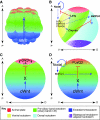
A four-step model for specification and organization of the sea urchin embryo nervous system. (A) In the first step, an anterior neuroectoderm regulatory state (red) is present throughout the egg and much of the embryo during early cleavage stages. (B) In the second step, which occurs during very early blastula stages, this state is eliminated by canonical Wnt (cWnt)-dependent signals from all but the anterior neuroectoderm, revealing a ciliary band-like neuroectoderm (green) that contains scattered neural precursors (light pink circles). (C) In the third step, which occurs during the mesenchyme blastula/early gastrula stages, Nodal and BMP2/4 signals convert ventral and dorsal ectoderm to non-neural ectoderm except in the anterior neuroectoderm (red) and ciliary band (green), which are protected from these signals. (D) During the fourth and final step, by which point the embryo has transitioned into a larva, CBE and ANE neural progenitors differentiate. The timeline indicates hours post-fertilization and embryonic stages.
None

Similar articles

Cited by

References

    1. Angerer L. M., Oleksyn D. W., Logan C. Y., McClay D. R., Dale L., Angerer R. C. (2000). A BMP pathway regulates cell fate allocation along the sea urchin animal-vegetal embryonic axis. Development 127, 1105-1114 - PubMed
    1. Angerer L. M., Oleksyn D. W., Levine A. M., Li X., Klein W. H., Angerer R. C. (2001). Sea urchin goosecoid function links fate specification along the animal-vegetal and oral-aboral embryonic axes. Development 128, 4393-4404 - PubMed
    1. Arendt D., Denes A. S., Jekely G., Tessmar-Raible K. (2008). The evolution of nervous system centralization. Philos. Trans. R. Soc. Lond. B Biol. Sci. 363, 1523-1528 - PMC - PubMed
    1. Bergeron K. F., Xu X., Brandhorst B. P. (2010). Oral-aboral patterning and gastrulation of sea urchin embryos depend on sulfated glycosaminoglycans. Mech. Dev. 128, 71-89 - PubMed
    1. Bisgrove B. W., Burke R. D. (1986). Development of serotongic neurons in embryos of the sea urchin, Strongylocentrotus purpuratus. Dev. Growth Differ. 28, 569-574 - PubMed

Publication types

MeSH terms

LinkOut - more resources