. 2011 Sep;138(17):3639-46.
doi: 10.1242/dev.067595.

An absolute requirement for Pax7-positive satellite cells in acute injury-induced skeletal muscle regeneration

Affiliations

An absolute requirement for Pax7-positive satellite cells in acute injury-induced skeletal muscle regeneration

Christoph Lepper et al. Development. 2011 Sep.

Abstract

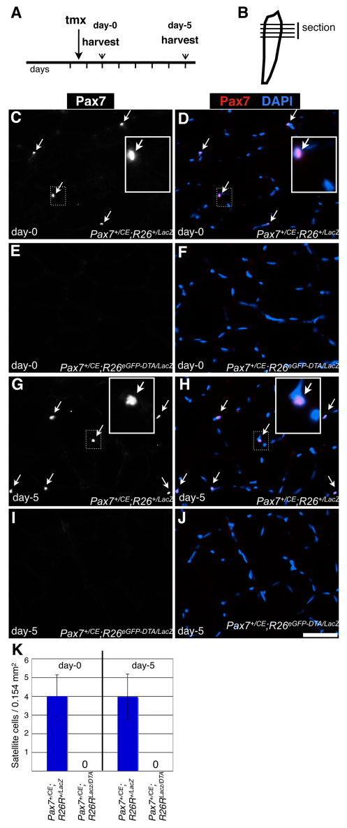
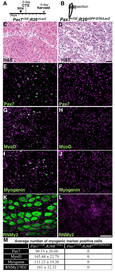
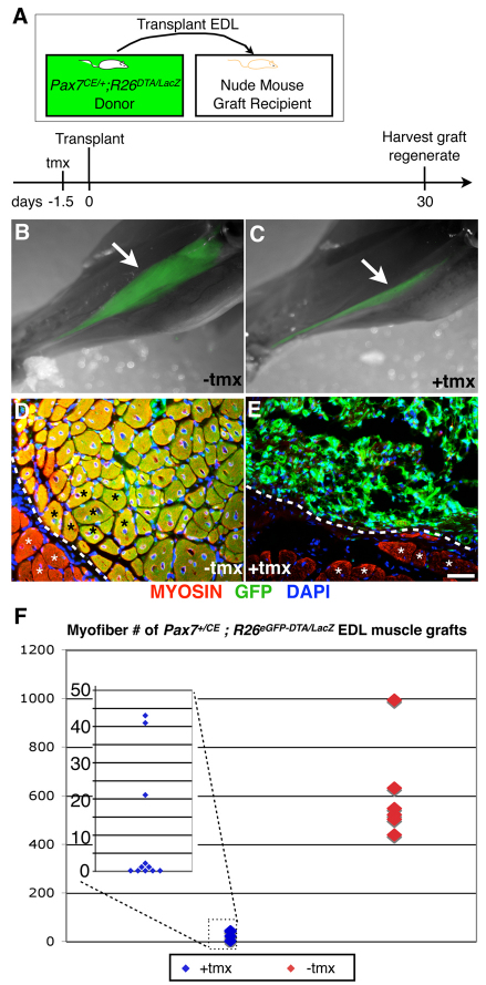
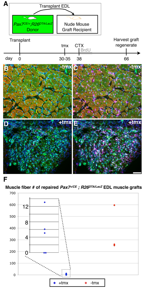
Skeletal muscle tissue provides mechanical force for locomotion of all vertebrate animals. It is prone to damage from acute physical trauma and physiological stress. To cope with this, it possesses a tremendous capacity for rapid and effective repair that is widely held to be accomplished by the satellite cells lying between the muscle fiber plasmalemma and the basement membrane. Cell transplantation and lineage-tracing studies have demonstrated that Pax7-expressing (Pax7(+)) satellite cells can repair damaged muscle tissue repeatedly after several bouts of acute injury. These findings provided evidence that Pax7(+) cells are muscle stem cells. However, stem cells from a variety of other origins are also reported to contribute to myofibers upon engraftment into muscles, questioning whether satellite cells are the only stem cell source for muscle regeneration. Here, we have engineered genetic ablation of Pax7(+) cells to test whether there is any significant contribution to muscle regeneration after acute injury from cells other than this source. We find that such elimination of Pax7(+) cells completely blocks regenerative myogenesis either following injury to the tibialis anterior (TA) muscle or after transplantation of extensor digitorum longus (EDL) muscles into nude mice. As Pax7 is specifically expressed in satellite cells, we conclude that they are essential for acute injury-induced muscle regeneration. It remains to be established whether there is any significant role for stem cells of other origins. The implications of our results for muscle stem cell-based therapy are discussed.


Figures

Fig. 1.
Fig. 1.
Efficient ablation of Pax7+ cells. (A) Tamoxifen (tmx) regimen and harvest scheme. Vertical lines indicate daily intervals. (B) Tibialis anterior (TA) muscle diagram. Horizontal lines indicate the orientation of sections (i.e. cross-sections) used for analysis. (C-J) Fluorescent microscopy of Pax7+/CE;R26R+/lacz (C,D,G,H) and Pax7+/CE;R26ReGFP-DTA/lacz (E,F,I,J) TA muscle cross-sections at 1.5 days after tmx administration (C-F) [referred to as day-0 as cardiotoxin (CTX)-induced injuries are performed at this time (see Fig. 2)] and 6.5 days after tamoxifen administration (G-J) [referred to as day-5 as this represents the 5-day regeneration time point (see Fig. 2)]. Pax7 is shown in white (C,E,G,I) or pseudocolored in magenta with DAPI shown in cyan (D,F,H,J). White arrows indicate satellite cells. Boxed insets show magnified views of the area indicated by a dotted white box. (K) Mean number of satellite cells at day-0 (left) and day-5 (right) per 0.154 mm2; 20 random areas/animal (n=3/genotype). Error bars represent s.d. Scale bar: 50 μm.
Fig. 2.
Fig. 2.
Absence of the myogenic response to acute injury in satellite cell-ablated mice. (A) Tamoxifen (tmx) and cardiotoxin (CTX) regimen and harvest scheme. Vertical lines indicate daily intervals. (B) Tibialis anterior (TA) muscle diagram with injury in gray. Horizontal lines indicate cross-sections used for analysis. (C-L) Pax7+/CE;R26R+/lacz (C,E,G,I,K) and Pax7+/CE;R26ReGFP-DTA/lacz (D,F,H,J,L) regenerated TA muscle cross-sections 5 days after CTX-induced injury. (Note: injured area is shown exclusively.) (C,D) Hematoxylin and Eosin stain. (E-L) Fluorescent microscopy of Pax7 (E,F), MyoD (G,H), myogenin (I,J) and RNMy2/9D2 (K,L). All markers are pseudocolored in green, DAPI in magenta. Scale bars: in D, 50 μm for C,D; in L, 50 μm for E-L. (M) Average numbers (±s.d.) of cells expressing the indicated myogenic markers 5 days after injury (each average is based on three random 0.154 mm2 regenerate areas per animal, n=3 per genotype).
Fig. 3.
Fig. 3.
Absence of regenerative myofibers in extensor digitorum longus (EDL) muscle grafts ablated of Pax7+ cells prior to transplantation. (A) Tamoxifen (tmx) regimen, EDL transplantation and harvest scheme. (B,C) Overlays of whole-mount bright-field and GFP epi-fluorescence (green) images of lower hindlimb muscles from Pax7+/CE;R26ReGFP-DTA/lacz without (B) and with (C) exposure of the donor EDL muscle to tmx prior to transplantation. Note that EDL muscles exposed to tmx to kill Pax7+ cells prior to transplantation fail to generate robust muscle grafts. White arrows indicate grafted tissue. (D,E) Fluorescent microscopy on cross-sections of Pax7+/CE;R26ReGFP-DTA/lacz EDL grafts without (D) and with (E) tmx treatment prior to transplantation. Myosin is pseudocolored in red, GFP in green and DAPI in blue. The host-graft interface is demarcated by the white dotted line. White asterisks indicate host myofibers; black asterisks indicate graft myofibers. Scale bar: 50 μm. (F) Comparison of total myofiber numbers of EDL grafts shows a clear difference between grafts with (blue) and without (red) tamoxifen treatment prior to transplantation (P=0.0002, two-tailed Mann-Whitney U-test). As the data points for tmx-treated EDL muscles cluster closely together, all data points are shown in the magnified inset (overlapping data points are plotted next to each other, e.g. five data points for `0' myofibers).
Fig. 4.
Fig. 4.
Robust vascularization of EDL muscles transplanted with or without prior ablation of Pax7+ cells and innervation of control EDL muscle grafts. (A-F) Fluorescent microscopy of Pax7+/CE;R26ReGFP-DTA/lacz EDL grafts without (A,C,E,F) and with (B,D) exposure of the donor EDL muscle to tamoxifen (tmx) prior to transplantation. (A,B) PECAM is pseudocolored in red, GFP in green and DAPI in blue. White arrows indicate PECAM+ capillaries; white arrowheads indicate GFP/PECAM+ host infiltrating cells. (C,D) CD34 is pseudocolored in red, GFP in green and DAPI in blue. White arrows indicate CD34+ capillaries; white arrowheads indicate GFP/CD34+ host infiltrating cells. (E) 2H3 (neurofilament)-stained neuronal axons are pseudocolored in red, GFP in green and DAPI in blue. White arrows indicate 2H3+/GFP neuronal processes. (F) Alexa568-conjugated α-bungarotoxin-labeled motor end plates (via binding to nicotinic acetylcholine receptors) are pseudocolored in red, GFP in green and DAPI in blue. White arrows indicate bungarotoxin+/GFP motor end plates. Scale bar: 50 μm.
Fig. 5.
Fig. 5.
Absence of muscle regeneration potential in established EDL grafts ablated of Pax7+ cells. (A) EDL transplantation, tamoxifen (tmx) treatment, injury and harvest scheme. (B-E) Fluorescent microscopy on cross-sections of established Pax7+/CE;R26ReGFP-DTA/lacz EDL grafts four weeks post-cardiotoxin (CTX)-induced injury without (B,C) and with (D,E) tmx treatment one month after transplantation prior to injury. Myosin/GFP/DAPI overlay is shown in B and D; Myosin/GFP/BrdU/DAPI overlay in C and E. Myosin is pseudocolored in red, GFP in green, DAPI in blue and BrdU in magenta. White arrows indicate BrdU+ central myonuclei. Scale bar: 50 μm. (F) Comparison of total myofiber numbers of EDL grafts four weeks after injury shows a clear difference between muscles treated with (blue) and without (red) tmx treatment prior to injury (P=0.0357, two-tailed Mann-Whitney U-test). As the data points for EDL muscle regenerates after tmx-treatment cluster closely together, all data points are shown in the magnified inset (overlapping data points are plotted next to each other, e.g. two data points for `0' myofibers).

References

    1. Bigard A. X., Janmot C., Sanchez H., Serrurier B., Pollet S., d'Albis A. (1999). Changes in myosin heavy chain profile of mature regenerated muscle with endurance training in rat. Acta Physiol. Scand. 165, 185-192 - PubMed
    1. Bischoff R. (1975). Regeneration of single skeletal muscle fibers in vitro. Anat. Rec. 182, 215-235 - PubMed
    1. Cerletti M., Jurga S., Witczak C. A., Hirshman M. F., Shadrach J. L., Goodyear L. J., Wagers A. J. (2008). Highly efficient, functional engraftment of skeletal muscle stem cells in dystrophic muscles. Cell 134, 37-47 - PMC - PubMed
    1. Collins C. A., Olsen I., Zammit P. S., Heslop L., Petrie A., Partridge T. A., Morgan J. E. (2005). Stem cell function, self-renewal, and behavioral heterogeneity of cells from the adult muscle satellite cell niche. Cell 122, 289-301 - PubMed
    1. Day K., Shefer G., Richardson J. B., Enikolopov G., Yablonka-Reuveni Z. (2007). Nestin-GFP reporter expression defines the quiescent state of skeletal muscle satellite cells. Dev. Biol. 304, 246-259 - PMC - PubMed

Publication types

MeSH terms