Clinical Trial
. 2011 Nov;96(11):1627-35.
doi: 10.3324/haematol.2011.047993. Epub 2011 Aug 9.

High VLA-4 expression is associated with adverse outcome and distinct gene expression changes in childhood B-cell precursor acute lymphoblastic leukemia at first relapse

Affiliations
Clinical Trial

High VLA-4 expression is associated with adverse outcome and distinct gene expression changes in childhood B-cell precursor acute lymphoblastic leukemia at first relapse

Shabnam Shalapour et al. Haematologica. 2011 Nov.

Abstract

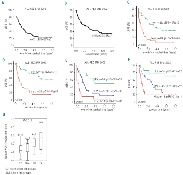
Background: Resistance to therapy and subsequent relapse remain major challenges in the clinical management of relapsed childhood acute lymphoblastic leukemia. As the bone marrow environment plays an important role in survival and chemotherapy resistance of leukemia cells by activating different signaling pathways, such as the VLA-4 and PI3K/Akt pathways, we studied the prognostic and biological impact of VLA-4 expression in leukemia cells from children with relapsed B-cell precursor acute lymphoblastic leukemia and its influence on the sensitivity of the leukemia cells to drugs.

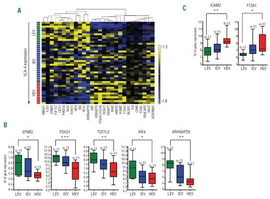
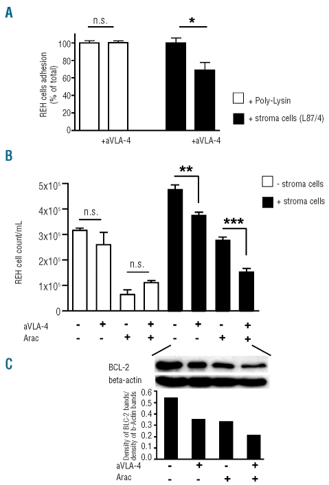
Design and methods: VLA-4 expression was quantified by real-time polymerase chain reaction in leukemia cells from 56 patients with relapsed acute lymphoblastic leukemia enrolled in the ALL-REZ BFM 2002 trial of the Berlin-Frankfurt-Münster study group. Gene expression changes related to VLA-4 expression were investigated by microarray-based mRNA profiling. The effect of VLA-4 signaling on proliferation and drug resistance was studied in co-cultures of leukemia and stromal cells.

Results: High expression of VLA-4 at first relapse was associated with adverse prognostic factors, poor molecular response to therapy and significantly worse probabilities of event-free and overall survival. VLA-4 expression was an independent prognostic parameter. Comparing gene expression profiles of leukemia cells with high versus low VLA-4 expression, we identified 27 differentially expressed genes primarily involved in the PI3K/Akt, ephrin and Rho GTPase pathways. Blocking of VLA-4 signaling in combination with cytarabine treatment abolished the growth supportive effect of stromal cells.

Conclusions: Our results show that high VLA-4 expression is a marker of poor prognosis and a potential therapeutic target in children with relapsed acute lymphoblastic leukemia and confirm that cellular interactions and biological effects related to VLA-4 play a decisive role in the survival of leukemia cells and response to therapy. (ClinicalTrials.gov identifier: NCT00114348).


Figures

Figure 1.
Figure 1.
VLA-4 expression is associated with outcome of BCP-ALL at first relapse. (A) Kaplan-Meier analysis of event-free survival (EFS) is shown for the total VLA-4 BCP-ALL patient cohort (n=51). (B) Kaplan-Meier analysis of overall survival (OS) is shown for the total VLA-4 BCP-ALL patient cohort (n=51). (C) Kaplan-Meier analysis of EFS is shown for BCP-ALL patients (n=51) with VLA-4 expression levels in leukemia cells lower and higher than the median (8.3). (D) Kaplan-Meier analysis of OS is shown for BCP-ALL patients (n=51) with VLA-4 expression levels in leukemia cells lower and higher than the median. (E) Kaplan-Meier analysis of EFS for three groups of BCP-ALL patients (n=51) with VLA-4 low (lower quartile, < 25%; LEV), intermediate (intermediate quartiles, >25%–<75%; IEV) and high expression level (upper quartile, >75%; HEV). (F) Kaplan-Meier analysis of OS for three groups of BCP-ALL patients (n=51) with VLA-4 low (LEV), intermediate (IEV) and high (HEV) expression level. (G) Comparison of VLA-4 expression levels between four different risk stratification groups of the ALL-REZ-BMF 2002 trial (Kruskal Wallis test: P=0.012). S2-: S2 group with MRD < 103 at day 36; S2+: S2 group with MRD ≥ 10−3 at day 36; S3 and S4, high-risk groups.
Figure 2.
Figure 2.
VLA-4 expression is associated with distinctive gene expression changes. (A) Expression levels of 27 genes identified by comparison of relapsed BCP-ALL patients with low (lower quartile, < 25%; LEV, green), intermediate (intermediate quartiles, >25%–<75%; IEV blue), and high (upper quartile, >75%; HEV, red) expression of VLA-4. Expression levels are shown as differences with respect to the mean over all patients (n=43). Patients were ordered according to VLA-4 expression levels. Genes are clustered according to down-and up-regulation in HEV- versus LEV-groups. (B) Box plots of down-regulated genes in HEV- versus LEV-groups obtained by chip analysis in the three groups (LEV: green; IEV: blue; HEV: red). Signal intensities (SI) of expression of five representative genes: EFNB2, FOXO1, TCF7L2, IRF4, and ARHGAP29 are shown. (C) Box plots of up-regulated genes in the HEV- versus LEV-groups, obtained by chip analysis in the three groups (LEV: green; IEV: blue; HEV: red). Signal intensities (SI) of expression of two representative genes are shown: ICAM2 and F13A1. Statistical analysis was done by ANOVA (*P<0.05; **P<0.001; ***P<0.0001).
Figure 3.
Figure 3.
Blocking of VLA-4 signaling overcomes the supportive effect of stromal cells. (A) For cell adhesion assay, the BCP-ALL cells (REH) were fluorescence-labeled and seeded into 24-well plates coated with poly-lysine or the stromal cell line L87/4 in the absence or presence of the blocking anti-VLA-4 monoclonal antibody. The percentage of adherent cells measured by the fluorescence plate reader is shown relative to control samples without blocking antibodies. (B) REH cells were cultured either alone or in co-culture with L87/4 stromal cells with or without blocking anti VLA-4 monoclonal antibody and cytarabine (ARA-C, 1 …M). After 48 h, proliferation was measured by the MTS tetrazolium assay and cell numbers per microliter were calculated from appropriate cell dilution series. Columns, mean of at least three independent experiments; bars, SD (**P<0.01; ***P<0.001). (C) Western-blot analysis of the anti-apoptotic protein BCL-2. REH cells were grown in co-culture with L87/4 stromal cells and were treated with or without blocking anti VLA-4 monoclonal antibody and/or ARA-C for 48 h. β-actin served as the control for equal protein loading. The density of dots was measured. The ratios of BCL-2 density to β-actin density are shown.

References

    1. Salzer WL, Devidas M, Carroll WL, Winick N, Pullen J, Hunger SP, et al. Long-term results of the pediatric oncology group studies for childhood acute lymphoblastic leukemia 1984–2001: a report from the children’s oncology group. Leukemia. 2010;24(2):355–70. - PMC - PubMed
    1. Moricke A, Zimmermann M, Reiter A, Henze G, Schrauder A, Gadner H, et al. Long-term results of five consecutive trials in childhood acute lymphoblastic leukemia performed by the ALL-BFM study group from 1981 to 2000. Leukemia. 2010;24(2):265–84. - PubMed
    1. Pui CH, Evans WE. Treatment of acute lymphoblastic leukemia. N Engl J Med. 2006;354(2):166–78. - PubMed
    1. Tallen G, Ratei R, Mann G, Kaspers G, Niggli F, Karachunsky A, et al. Long-term outcome in children with relapsed acute lymphoblastic leukemia after time-point and site-of-relapse stratification and intensified short-course multidrug chemotherapy: results of trial ALL-REZ BFM 90. J Clin Oncol. 2010;28(14):2339–47. - PubMed
    1. Eckert C, Biondi A, Seeger K, Cazzaniga G, Hartmann R, Beyermann B, et al. Prognostic value of minimal residual disease in relapsed childhood acute lymphoblastic leukaemia. Lancet. 2001;358(9289):1239–41. - PubMed

Publication types

MeSH terms

Supplementary concepts

Associated data