. 2011 Oct 20;118(16):4449-53.
doi: 10.1182/blood-2011-04-345876. Epub 2011 Aug 9.

Extracellular matrix structure and nano-mechanics determine megakaryocyte function

Affiliations

Extracellular matrix structure and nano-mechanics determine megakaryocyte function

Alessandro Malara et al. Blood. .

Abstract

Cell interactions with matrices via specific receptors control many functions, with chemistry, physics, and membrane elasticity as fundamental elements of the processes involved. Little is known about how biochemical and biophysical processes integrate to generate force and, ultimately, to regulate hemopoiesis into the bone marrow-matrix environment. To address this hypothesis, in this work we focus on the regulation of MK development by type I collagen. By atomic force microscopy analysis, we demonstrate that the tensile strength of fibrils in type I collagen structure is a fundamental requirement to regulate cytoskeleton contractility of human MKs through the activation of integrin-α2β1-dependent Rho-ROCK pathway and MLC-2 phosphorylation. Most importantly, this mechanism seemed to mediate MK migration, fibronectin assembly, and platelet formation. On the contrary, a decrease in mechanical tension caused by N-acetylation of lysine side chains in type I collagen completely reverted these processes by preventing fibrillogenesis.


Figures
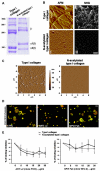
Figure 1
Figure 1
Characterization of collagen nano-mechanical properties. (A) Coomassie staining of type I collagen and N-acetylated collagen. A total of 2 μg of proteins was loaded on 6% gel of polyacrylamide; > 85% of lysine residue were modified in the native protein determining a different migration in electrophoresis. (B) In vitro analysis of collagens structure. (i) Atomic force microscopy images of 3 μm2 of dehydrated collagen coating. (ii) Second harmonic generation images of collagen supramolecular structure were acquired through a 63× (1.2 NA) objective on a Leica DMIRE2 microscope with a TCS SP2 scanner. Analysis was performed with the Leica confocal software and ImageJ (NIH). Scale bar represents 10 μm. (C) Atomic force microscopy images of collagens in fluid (PBS). A total of 5 μm2 fields were analyzed in contact mode using an MFP-3D Bio-atomic force microscope (Asylum Research). (D) Roles of α2-integrin and GPVI on MK adhesion and spreading on type I collagen. Cord blood-derived MKs were seeded for 2 hours on type I collagen in the presence of 20 μg/mL of anti-α2 (clone P1E6) and Fab GPVI (clone 9012.2) antibodies, fixed, and then stained for actin (red) and CD41 (green). Images were acquired through an Olympus BX51 using a 20×/0.5 UPlan objective. Scale bar represents 50 μm. (E) Effect of chemical modification of type I collagen on α2 integrin and GPVI engagement. MKs were seeded on type I collagen and N-acetylated type I collagen for 1 hour, and cell adhesion was evaluated in the presence of increasing concentrations of anti-α2 and GPVI antibodies. Cell adhesion values are expressed relative to control (absence of inhibitor).
Figure 2
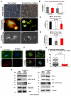
Figure 2
Effects of collagen nano-mechanical properties on MK behavior. (Ai-ii) Phase-contrast images of MKs seeded for 16 hours in adhesion on collagens. Scale bar represents 100 μm. (iii-iv) Stress fiber formation was analyzed after actin (tetramethylrhodamine isothiocyanate [TRITC]-phalloidin, red) staining in immunofluorescence, whereas proplatelet formation was evaluated using an anti–α-tubulin (green) antibody. Nuclei were counterstained with Hoechst 33288 (blue). Images were acquired using a 63×/1.25 UPlanF1 oil-immersion objective. Scale bar represents 20 μm. (v-vi) Scanning electron micrographs of spread MKs on type I collagen and a platelet-releasing MK on N-acetylated type I collagen. Images were acquired with a Cambridge Stereoscan 440 microscope (Leica Microsystems) at 17.5 kV and a magnification of 2000×. Scale bar represents 4 μm. (B) Evaluation of cell spreading after 2 and 16 hours of adhesion. Cells were fixed and stained with TRITC-phallodin. Cells exhibiting stress fibers were counted and presented as mean ± SD of 3 different experiments. P < .05. SHG indicates second harmonic generation. (C) Proplatelet formation was analyzed after 16-hour adhesion on different collagens or in MKs maintained in suspension (none). P < .05. (D) Migration of MKs in transwell plate after 16 hours of incubation; 8-μm polycarbonate pore filters were coated with native and N-acetylated collagen or with BSA (none), and cells were counted after CD41 staining in immunofluorescence. Results are reported as percentage of migrated cells relative to control of 3 different experiments ± SD. (Ei-ii) Assembly of FN under static conditions. MKs were incubated with 25 μg/mL of FITC-labeled FN and then seeded on different matrices. FN fibrillogenesis was then visualized in immunofluorescence using a 63×/1.25 UPlanF1 oil-immersion objective. Hoechst 33288 was used for nuclei staining (blue). Scale bar represents 10 μm. (eiii-iv) Alternatively, cells were removed using deoxycholic acid (DOC) and the underlying matrix stained with an anti-FN antibody (red) and visualized in immunofluorescence, using a 40×/0.75 oil-immersion objective. Scale bar represents 20 μm. (F) Relocation of endogenous FN in MKs after 2- (left panels) and 16-hour (right panels) adhesion on collagens. Cells were stained for FN (red), α-tubulin (green), and Hoechst for nuclei (blue) using a 63×/1.25 UPlan oil-immersion objective. Scale bar represents 10 μm. (G) Distribution of Young modulus values of MKs in adhesion on different collagen samples. Three different experiments were performed, and at least 5 cells for sample were analyzed. P < .05. (H) Analysis of α2β1 and GPVI-dependent pathways in adhering MKs. Rho guanosine triphosphate pull-down experiments and immunoblot analysis of endogenous MLC2, Syk, and Src phosphorylation levels in MKs adhering to native and N-acetylated type I collagen after 16-hour incubation, representative of 3 different experiments. Actin, tubulin, and nonmuscle myosin IIA were revealed to demonstrate equal protein loading.xxxxx.

References

    1. Shoulders MD, Raines RT. Collagen structure and stability. Annu Rev Biochem. 2009;78:929–958. - PMC - PubMed
    1. Chen Z, Naveiras O, Balduini A, et al. The May-Hegglin anomaly gene MYH9 is a negative regulator of platelet biogenesis modulated by the Rho-ROCK pathway. Blood. 2007;110(1):171–179. - PMC - PubMed
    1. Chang Y, Aurade F, Larbret F, et al. Proplatelet formation is regulated by the Rho/ROCK pathway. Blood. 2007;109(10):4229–4236. - PubMed
    1. Pallotta I, Lovett M, Rice W, et al. Bone marrow osteoblastic niche: a new model to study physiological regulation of megakaryopoiesis. PLoS One. 2009;4(12):e8359. - PMC - PubMed
    1. Spurlin TA, Bhadriraju K, Chung KH, et al. The treatment of collagen fibrils by tissue transglutaminase to promote vascular smooth muscle cell contractile signaling. Biomaterials. 2009;30(29):5486–5496. - PubMed

Publication types