. 2011 Sep 29;118(13):3559-69.
doi: 10.1182/blood-2011-06-357996. Epub 2011 Aug 9.

DNA methyltransferase 1 and DNA methylation patterning contribute to germinal center B-cell differentiation

Affiliations

DNA methyltransferase 1 and DNA methylation patterning contribute to germinal center B-cell differentiation

Rita Shaknovich et al. Blood. .

Abstract

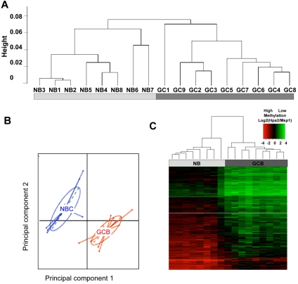
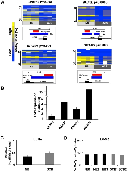
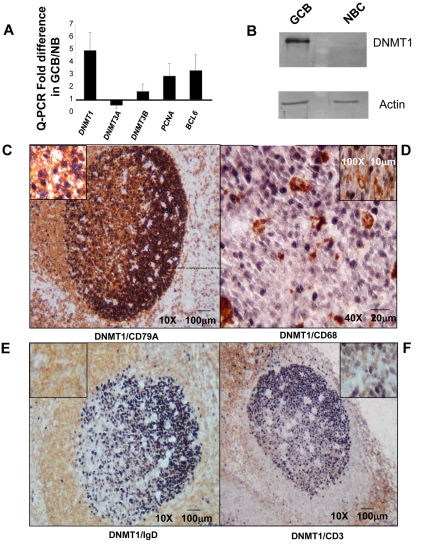
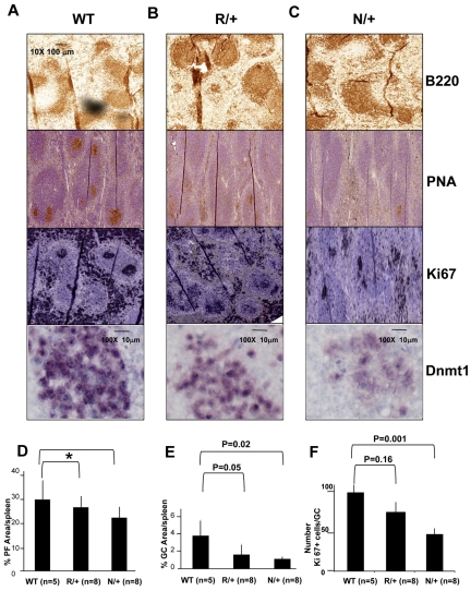
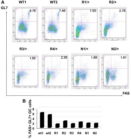
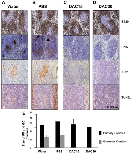
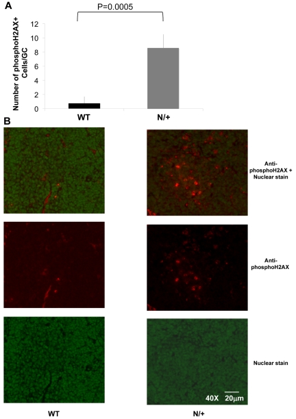
The phenotype of germinal center (GC) B cells includes the unique ability to tolerate rapid proliferation and the mutagenic actions of activation induced cytosine deaminase (AICDA). Given the importance of epigenetic patterning in determining cellular phenotypes, we examined DNA methylation and the role of DNA methyltransferases in the formation of GCs. DNA methylation profiling revealed a marked shift in DNA methylation patterning in GC B cells versus resting/naive B cells. This shift included significant differential methylation of 235 genes, with concordant inverse changes in gene expression affecting most notably genes of the NFkB and MAP kinase signaling pathways. GC B cells were predominantly hypomethylated compared with naive B cells and AICDA binding sites were highly overrepresented among hypomethylated loci. GC B cells also exhibited greater DNA methylation heterogeneity than naive B cells. Among DNA methyltransferases (DNMTs), only DNMT1 was significantly up-regulated in GC B cells. Dnmt1 hypomorphic mice displayed deficient GC formation and treatment of mice with the DNA methyltransferase inhibitor decitabine resulted in failure to form GCs after immune stimulation. Notably, the GC B cells of Dnmt1 hypomorphic animals showed evidence of increased DNA damage, suggesting dual roles for DNMT1 in DNA methylation and double strand DNA break repair.


Figures

Figure 1
Figure 1
GC B-cells feature a predominantly hypomethylated DNA methylation signature. (A) Unsupervised hierarchical clustering using the Ward method was performed on all probesets and accurately segregated NB cells from GC B cells (GCB). (B) Principal component analysis of methylation values for NBs and GCBs. The first and second principal components separate NBs from GCBs, underscoring the overall differences in methylation patterning. (C) A signature of differentially methylated genes in GC B-cells versus NB cells based on P < .01 (moderated t test with BH correction) and methylation difference of ∼ 40% was identified and included 235 genes. A heatmap representation allows visualization of the finding that the majority of differentially methylated genes are hypomethylated in GC B cells
Figure 2
Figure 2
Hypomethylation preferentially affects regulatory regions of the genome in GC B cells. (A) Four genes from the GC B-cell signature were selected for validation by MassArray. The results are represented as heatmaps in which the columns correspond to individual samples, while rows represent individual CpGs with color reflecting methylation value. P values are from moderated t test comparing methylation values from all tested CpGs between GC B cells and NBs. The location of MassArray amplicons (blue) and HELP probesets (red) relative to the transcriptional start site (TSS) of each gene (black) is illustrated below each heatmap. (B) The relative transcript abundance of the same 4 genes was measured by QPCR in 3 additional NB and GC B-cell specimens. The y-axis depicts fold expression difference in GC B-cells versus NBs calculated using ddCT method. All 4 genes are expressed at higher levels in GC B-cells, concordant with their hypomethylation. (C) LUMA assays performed on 3 NB and 2 GC B-cell specimens show a mild increase in the abundance of hypomethylated HpaII sites. The y-axis depicts relative signal of HpaII vs MspI signals. (D) Liquid chromatography-mass spectrometry was performed in 3 NB and 2 GC B-cell specimens. The y-axis depicts the percentage of methylcytosine versus total cytosines in each specimen.
Figure 3
Figure 3
DNMT1 is highly expressed in GC B cells. (A) Relative transcript abundance of DNMT1, DNMT3A, and DNMT3B as well as the GC B-cell specific transcripts PCNA and BCL6 were measured by QPCR. The y-axis depicts the fold difference of relative expression of each transcript in GC B cells versus NB cells. (B) Immunoblotting with anti-DNMT1 antibody was performed in GC B cells and NB cells as indicated, using actin as loading control. (C-F) Immunohistochemical double-staining in reactive human tonsillar tissue delineates expression of DNMT1 (dark purple nuclear staining), (C) in the germinal center CD79A+ (membranous brown staining) B cells and not in (D) CD68+ (membranous brown staining) tingible-body macrophages or (E) IgD+ (membranous brown staining) NB cells, or (F) CD3+ (membranous brown staining) T cells.
Figure 4
Figure 4
GC formation is impaired in Dnmt1 hypomorphic mice. (A-C) C57BL/6 wild type (WT; A), or Dnmt1 R/+ (B) and Dnmt1 N/+ (C) were immunized with SRBC to induce GC formation. Animals were killed on day 10 and formalin fixed paraffin embedded splenic tissue was stained with anti-B220, PNA, anti-Ki67, and anti-Dnmt1. (D) Quantitative imaging of spleen tissue based on immunohistochemistry staining from Figure 3A revealed no significant difference in the size of primary follicles (*P = .18 and .52, t test), but decreased size of the GCs (E) and the number of proliferating cells per GC (ki67) in the Dnmt1 hypomorph animals (F). The numbers of animals per condition are shown in parenthesis.
Figure 5
Figure 5
Reduction in the number of GC B cells in immunized Dnmt1 hypomorphic mice. (A) Mononuclear splenocytes were purified 10 days after immunization with SRBC from WT, Dnmt1R/+ and Dnmt1N/+ mice and stained with 7-AAD/ B220/GL7/FAS. The percentage of GL7+ FAS+ cells reflects the abundance of GC B cells in the various animals. (B) The results from panel A are represented as a histogram depicting the percentage of GC B cells for each of the mice analyzed by flow cytometry.
Figure 6
Figure 6
Decitabine treatment abrogates formation of GCs in mice. Twenty C57/BL6 mice were immunized with SRBCs and subjected to daily intraperitoneal injections of water (A), PBS solution (ie, vehicle, B), 15 mg/m2 decitabine (C) or 30 mg/m2 decitabine (D). Formalin fixed paraffin embedded splenic tissue recovered at day 10 was examined by immunohistochemistry for B220, PNA, Ki67 and TUNEL. (E) The histogram represents the quantitative assessment of primary follicle and GC size as assessed by immunohistochemistry staining in all animals.
Figure 7
Figure 7
Increased H2AX phosphorylation in the GCs of Dnmt1 hypomorphic mice. (A) The number of phosphoH2AX positive cells per GC was counted in WT or Dnmt1N/+ mice at day 10 after immunization with SRBC based on immunofluorescent staining of splenic sections (B) Representative images from the immunofluorescent staining of WT and Dnmt1N/+ hypomorphic mice using a nuclear stain (green) and anti-phosphoH2AX monoclonal antibody (red), (top: overlay; middle: anti-phosphoH2AX antibody; and bottom: nuclear stain only).

References

    1. Ci W, Polo JM, Melnick A. B-cell lymphoma 6 and the molecular pathogenesis of diffuse large B-cell lymphoma. Curr Opin Hematol. 2008;15(4):381–390. - PMC - PubMed
    1. Klein U, Dalla-Favera R. Germinal centres: role in B-cell physiology and malignancy. Nat Rev Immunol. 2008;8(1):22–33. - PubMed
    1. de Yebenes VG, Ramiro AR. Activation-induced deaminase: light and dark sides. Trends Mol Med. 2006;12(9):432–439. - PubMed
    1. Martin A, Bardwell PD, Woo CJ, Fan M, Shulman MJ, Scharff MD. Activation-induced cytidine deaminase turns on somatic hypermutation in hybridomas. Nature. 2002;415(6873):802–806. - PubMed
    1. Yamane A, Resch W, Kuo N, et al. Deep-sequencing identification of the genomic targets of the cytidine deaminase AID and its cofactor RPA in B lymphocytes. Nat Immunol. 2011;12(1):62–69. - PMC - PubMed

Publication types

MeSH terms

Substances