. 2011 Aug 9;2(4):e00167-11.
doi: 10.1128/mBio.00167-11. Print 2011.

Nonlytic exocytosis of Cryptococcus neoformans from macrophages occurs in vivo and is influenced by phagosomal pH

Affiliations

Nonlytic exocytosis of Cryptococcus neoformans from macrophages occurs in vivo and is influenced by phagosomal pH

André Moraes Nicola et al. mBio. .

Abstract

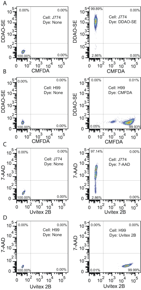
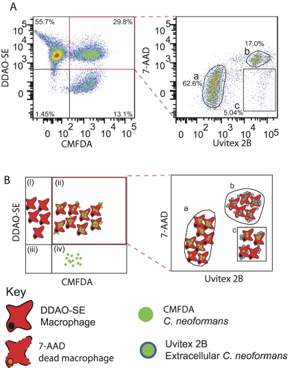
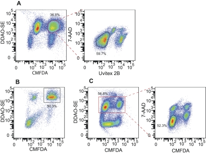
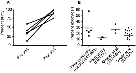
A unique aspect of the interaction of the fungus Cryptococcus neoformans with macrophages is the phenomenon of nonlytic exocytosis, also referred to as "vomocytosis" or phagosome extrusion/expulsion, which involves the escape of fungal cells from the phagocyte with the survival of both cell types. This phenomenon has been observed only in vitro using subjective and time-consuming microscopic techniques. In spite of recent advances in our knowledge about its mechanisms, a major question still remaining is whether this phenomenon also occurs in vivo. In this study, we describe a novel flow cytometric method that resulted in a substantial gain in throughput for studying phagocytosis and nonlytic exocytosis in vitro and used it to explore the occurrence of this phenomenon in a mouse model of infection. Furthermore, we tested the hypothesis that host cell phagosomal pH affected nonlytic exocytosis. The addition of the weak bases ammonium chloride and chloroquine resulted in a significant increase of nonlytic exocytosis events, whereas the vacuolar ATPase inhibitor bafilomycin A1 had the opposite effect. Although all three agents are known to neutralize phagosomal acidity, their disparate effects suggest that phagosomal pH is an important and complex variable in this process. Our experiments established that nonlytic exocytosis occurred in vivo with a frequency that is possibly much higher than that observed in vitro. These results in turn suggest that nonlytic exocytosis has a potential role in the pathogenesis of cryptococcosis.

Importance: Cryptococcus neoformans causes disease in people with immune deficiencies such as AIDS. Upon infection, C. neoformans cells are ingested by macrophage immune cells, which provide a niche for survival and replication. After ingestion, macrophages can expel the fungi without causing harm to either cell type, a process named nonlytic exocytosis. To dissect this phenomenon, we evaluated its dependence on the pH inside the macrophage and addressed its occurrence during infection of mice. We developed new techniques using flow cytometry to measure C. neoformans internalization by and nonlytic exocytosis from macrophages. Neutralizing the phagosome acidity changed the rate of nonlytic exocytosis: activity increased with the weak bases chloroquine and ammonium chloride, whereas the vacuolar ATPase inhibitor bafilomycin A1 caused it to decrease. Experiments in mice suggested that nonlytic exocytosis occurred during infection with C. neoformans. These results shed new light on the interaction between C. neoformans and host macrophages.


Figures

FIG 1
FIG 1
Flow cytometry analysis of J774 and C. neoformans cells stained with different fluorescent dyes. J774 cells were stained with either DDAO-SE (A) or 7-AAD (C), and C. neoformans strain H99 was stained with either CMFDA (B) or Uvitex 2B (D). Unstained controls were included in each assay and are shown in the left column. The 7-AAD-stained J774 cells had been previously fixed with 1% paraformaldehyde and permeabilized with 0.1% saponin to mimic dead cells. This experiment was repeated at least twice with similar results.
FIG 2
FIG 2
Flow cytometry phagocytosis assay. (A) Flow cytometric analysis of phagocytosis after 2 h of coincubation. (B) Diagrammatic representation of the plots in panel A. C. neoformans and J774 cells were stained with CMFDA (green) and DDAO-SE (red), respectively; phagocytosed for 2 h; and stained with Uvitex 2B (blue) and 7-AAD (orange) prior to flow cytometry. The density plot on the left shows macrophages (i), macrophages associated with C. neoformans (ii), debris (iii), and C. neoformans cells (iv). Events in the double-positive gate (highlighted in red) were plotted for Uvitex 2B and 7-AAD fluorescence intensity (right plot) to identify internalized C. neoformans and determine phagocyte viability. Three populations are distinguishable: (a) Uvitex 2B-negative, 7-AAD-negative, live macrophages with internalized C. neoformans; (b) 7-AAD-positive, dead phagocytes; and (c) Uvitex 2B-positive, 7-AAD-negative, live phagocytes with attached C. neoformans. This experiment was repeated at least 10 times with similar results. Note that the right plot in panel A does not have a clearly defined 7-AAD-positive, Uvitex 2B-negative population. This is not a compensation artifact (see Fig. S8 in the supplemental material) but is due to the fact that the membrane rupture of dead macrophages that permits penetration of 7-AAD also permits penetration of Uvitex 2B, which then labels internalized in addition to attached C. neoformans.
FIG 3
FIG 3
Quantification of nonlytic exocytosis using flow cytometry. Stained cells were allowed to phagocytose for 2 h and analyzed in three steps of flow cytometry to determine nonlytic exocytosis rates. (A) Events to be sorted were gated for forward and side scatter followed by gating as positive for CMFDA and DDAO-SE (highlighted in red) and negative for Uvitex 2B and 7-AAD. (B) Cells immediately postsorting, demonstrating enrichment of macrophages with internalized C. neoformans (gate). (C) Sorted cells were incubated for 24 h and analyzed again. Nonlytic exocytosis events were defined by gating as DDAO-SE positive (highlighted in red) and CMFDA negative, 7-AAD negative. This experiment was repeated at least 10 times with similar results.
FIG 4
FIG 4
Comparison between microscopic and flow cytometric measurements of nonlytic exocytosis. (A) Percentage of macrophages that have internalized C. neoformans cells (DDAO-SE positive/CMFDA positive/Uvitex 2B negative divided by total DDAO-SE positive) prior to and immediately after sorting. Results are from five independent experiments done on different days. (B) Distribution of percent nonlytic exocytosis measured in each individual replicate by time-lapse imaging and flow cytometry. The numbers in parentheses represent the macrophages that exocytosed and the total number of macrophages observed when all replicates were pooled. Data in the last two columns come from previously published results using time-lapse microscopy (14, 25) and have been replotted for comparison with the flow cytometry method. The results reported by Voelz et al. reflect nonlytic exocytosis measured for 20 h in serum-free medium, whereas all other experiments were done in serum-containing medium for 24 h. Bars represent means; each condition was tested at least three times on different days.
FIG 5
FIG 5
Evidence for nonlytic exocytosis in vivo. BMM were prepared for sorting as described for J774 cells. See Fig. S7 in the supplemental material for an outline of the experiment’s rationale. (A) In vitro assay. (B) Sorted stained cells that were injected intratracheally into mice and harvested by bronchoalveolar lavage 24 h later for analysis. (C) Unstained cells that were injected, collected, and analyzed as described for panel B. This experiment was repeated twice on different days. The reader should refer to Fig. S7, which includes a schematic diagram of the experiment detailing the various steps and provides cartoons to assist in the interpretation of these flow cytometry data.

References

    1. Casadevall A, Perfect JR. 1998. Cryptococcus neoformans, 1st ed. ASM Press, Washington, DC
    1. Park BJ, et al. 2009. Estimation of the current global burden of cryptococcal meningitis among persons living with HIV/AIDS. AIDS 23:525–530 - PubMed
    1. Feldmesser M, Kress Y, Novikoff P, Casadevall A. 2000. Cryptococcus neoformans is a facultative intracellular pathogen in murine pulmonary infection. Infect. Immun. 68:4225–4237 - PMC - PubMed
    1. Shao X, et al. 2005. An innate immune system cell is a major determinant of species-related susceptibility differences to fungal pneumonia. J. Immunol. 175:3244–3251 - PubMed
    1. Zaragoza O, Alvarez M, Telzak A, Rivera J, Casadevall A. 2007. The relative susceptibility of mouse strains to pulmonary Cryptococcus neoformans infection is associated with pleiotropic differences in the immune response. Infect. Immun. 75:2729–2739 - PMC - PubMed

Publication types

LinkOut - more resources