

doi: 10.1101/gad.16750211.
Tsc1 mutant neural stem/progenitor cells exhibit migration deficits and give rise to subependymal lesions in the lateral ventricle
Affiliations
- PMID: 21828270
- PMCID: PMC3182017
- DOI: 10.1101/gad.16750211
Item in Clipboard
Tsc1 mutant neural stem/progenitor cells exhibit migration deficits and give rise to subependymal lesions in the lateral ventricle
Genes Dev.
.
Abstract
Subependymal nodules (SENs) and subependymal giant cell astrocytomas (SEGAs) are common brain lesions found in patients with tuberous sclerosis complex (TSC). These brain lesions present a mixed glioneuronal phenotype and have been hypothesized to originate from neural stem cells. However, this hypothesis has not been tested empirically. Here, we report that loss of Tsc1 in mouse subventricular zone (SVZ) neural stem/progenitor cells (NSPCs) results in formation of SEN- and SEGA-like structural abnormalities in the lateral ventricle, the consequence of abnormal migration of NSPCs following Tsc1 loss.
Figures
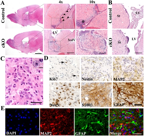
Tsc1Nestin cKO mice develop SEN- and SEGA-like abnormalities. (A) Tsc1Nestin cKO mice develop hydrocephalus and small tumors in the lateral ventricle, as shown by H&E staining on mice induced at P7 and examined at 3 mo of age. (Arrowheads) Interventricular foramen (IF); (*) stem cell-rich subventricular region; (LV) lateral ventricle; (3rdV) third ventricle. Bars: for whole forebrain pictures, 2 mm; for 4× pictures, 500 μm; for 10× pictures, 200 μm. (B) Tsc1Nestin cKO mice develop small nodular excrescences in the lateral ventricle (mice induced at P7, examined at 3 mo of age). (St) Striatum. Bar, 200 μm. (C) Histological features of the inner tumor cells near the IF. Bar, 40 μm. Inset shows a giant cell within the tumor mass. Bar, 10 μm. (D) Inner tumor cells are mostly cells immunoreactive for either neuronal (MAP2 and NeuN) or astrocytic (S100β and GFAP) markers. (Arrows) Ki67+ cells. Bar, 50 μm. (E) MAP2 (red) and GFAP (green) double-staining reveals mixed glioneuronal phenotype of cells inside the tumor and their fibrillated processes. Bar, 50 μm.
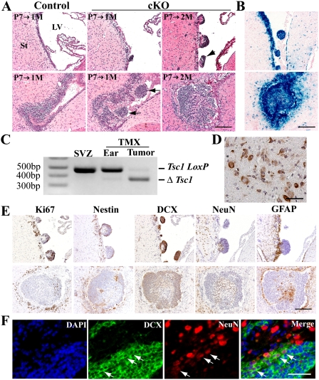
Development of small nodular structures and tumors in the lateral ventricle involves aberrant aggregation and migration of SVZ NSPCs. (A) Tsc1Nestin cKO mice progressively develop abnormalities in the lateral ventricle, as shown by mice induced at P7 and examined at 1 and 2 mo of age. (Arrows) Clusters of cells show aberrant aggregation and migration from the SVZ to the lateral ventricle. Bar, 200 μm. (B) Both nodular structures and early tumor masses are composed of cells in which Cre activity has been induced, as visualized by X-gal staining on brain sections of Tsc1Nestin cKO mice bearing the R26-lacZ reporter allele. Bar, 200 μm. (C) PCR assay detected the recombined Tsc1 allele (ΔTsc1 allele) in tumor samples but not in controls (SVZ samples of untreated Nestin-CreERT2;Tsc1loxp/loxp mice and ear samples of TMX-treated Nestin-CreERT2;Tsc1loxp/loxp). Tsc1 loxP is the flox allele. (D) Phospho-S6 staining indicates strong mTOR signaling in the tumor mass. Bar, 50 μm. (E) Nodular structures and tumors that develop in the ventricle originate from NSPCs. Bar, 200 μm. (F) Neuroblasts retained in the lateral ventricle give rise to neurons, as shown by DCX (green) and NeuN (red) double-staining. (Arrows) Cells that are double-positive for NeuNdim and DCX. Bar, 50 μm.

Tsc1Nestin cKO mice have fewer granule cells in the OB due to reduced migration. (A) Tsc1Nestin cKO mice have less granule cells in the OB. Bars: left panels, 500 μm; right panels, 100 μm. (B) Quantification of cell density in medial granule cell layer (GCL) of OB. n = 3 mice were examined for each genotype. Data are plotted by mean ± SEM and analyzed by Student's t-test; (**) P < 0.01. (C) Fewer newly generated granule cells in the OB were observed in Tsc1Nestin cKO mice. Bar, 50 bar. (D) Quantification of β-gal-positive cells in the medial granule cell layer. n = 3 pairs of mice were examined. Data are plotted by mean ± SEM and analyzed by Student's t-test; (**) P < 0.01.

Tsc1 loss in transit-amplifying neural progenitors results in a neuroblast migration deficit. (A) Cre activity was induced in both SVZ NSPCs and ependymal cells in Nestin-CreERT2 mice, while in the Ascl1-CreERTM mice, only a limited number of SVZ progenitor cells are targeted upon TMX induction. Ependymal cells are marked by S100β (red) staining. Cre activity is marked by YFP (green) staining. Nestin-CreERT2;Tsc1loxp/+;R26-YFP mice and Ascl1-CreERTM; Tsc1loxp/+;R26-YFP mice were treated with TMX at P7 (for Nestin-CreERT2 mice) or at P7, P9, and P11 (for Ascl1-CreERTM mice) and examined at 1 mo of age. Bar, 50 μm. (B) Diagram illustrating Ascl1 expression in transit-amplifying neural progenitor cells. (C) Gross examination revealed that Tsc1Ascl1 cKO mice develop small abnormal growths in the lateral ventricle near the IF. Bars: for whole forebrain pictures, 2 mm; for 4× pictures, 500 μm; for 10× pictures, 200 μm. (D) Abnormal growths in the lateral ventricle of Tsc1Ascl1 cKO mice are composed of Cre-positive cells. Cre activity is visualized by X-gal staining on brain sections of Tsc1AsclI heterozygous and cKO mice bearing the R26-lacZ reporter allele. Bar, 200 μm.
References
-
- Buccoliero AM, Franchi A, Castiglione F, Gheri CF, Mussa F, Giordano F, Genitori L, Taddei GL 2009. Subependymal giant cell astrocytoma (SEGA): is it an astrocytoma? Morphological, immunohistochemical and ultrastructural study. Neuropathology 29: 25–30 - PubMed
-
- Burger PC, Scheithauer BW, Vogel FS 2002. Surgical pathology of the nervous system and its coverings. Churchill Livingstone, New York.
-
- Ess KC, Kamp CA, Tu BP, Gutmann DH 2005. Developmental origin of subependymal giant cell astrocytoma in tuberous sclerosis complex. Neurology 64: 1446–1449 - PubMed
Publication types
MeSH terms
Substances
LinkOut - more resources
Full Text Sources
Molecular Biology Databases