. 2011 Aug 1;25(15):1628-40.
doi: 10.1101/gad.17269211.

An integrated approach to dissecting oncogene addiction implicates a Myb-coordinated self-renewal program as essential for leukemia maintenance

Affiliations

An integrated approach to dissecting oncogene addiction implicates a Myb-coordinated self-renewal program as essential for leukemia maintenance

Johannes Zuber et al. Genes Dev. .

Erratum in

  • Genes Dev. 2011 Sep 15;25(18):1997. Fellman, Christof [corrected to Fellmann, Christof]

Abstract

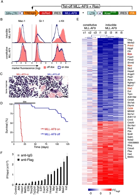
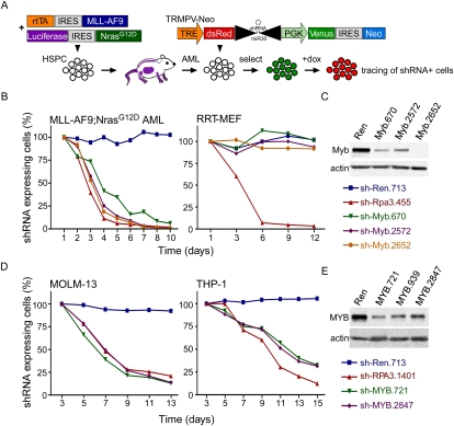
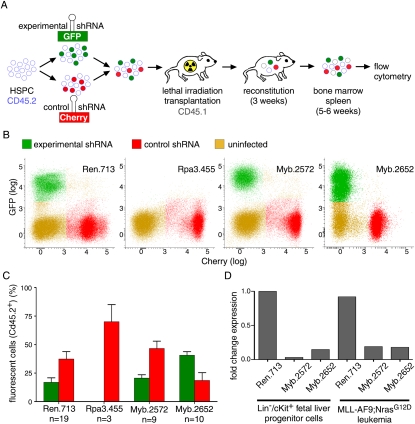
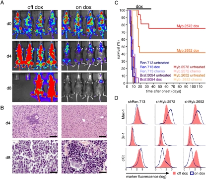
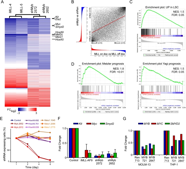
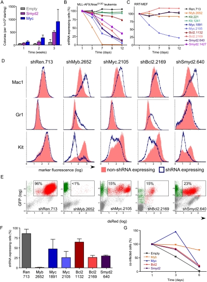
Although human cancers have complex genotypes and are genomically unstable, they often remain dependent on the continued presence of single-driver mutations-a phenomenon dubbed "oncogene addiction." Such dependencies have been demonstrated in mouse models, where conditional expression systems have revealed that oncogenes able to initiate cancer are often required for tumor maintenance and progression, thus validating the pathways they control as therapeutic targets. Here, we implement an integrative approach that combines genetically defined mouse models, transcriptional profiling, and a novel inducible RNAi platform to characterize cellular programs that underlie addiction to MLL-AF9-a fusion oncoprotein involved in aggressive forms of acute myeloid leukemia (AML). We show that MLL-AF9 contributes to leukemia maintenance by enforcing a Myb-coordinated program of aberrant self-renewal involving genes linked to leukemia stem cell potential and poor prognosis in human AML. Accordingly, partial and transient Myb suppression precisely phenocopies MLL-AF9 withdrawal and eradicates aggressive AML in vivo without preventing normal myelopoiesis, indicating that strategies to inhibit Myb-dependent aberrant self-renewal programs hold promise as effective and cancer-specific therapeutics. Together, our results identify Myb as a critical mediator of oncogene addiction in AML, delineate relevant Myb target genes that are amenable to pharmacologic inhibition, and establish a general approach for dissecting oncogene addiction in vivo.


Figures

Figure 1.
Figure 1.
Withdrawal of MLL-AF9 induces differentiation and disease remission. (A) Retroviral constructs used to generate AML. (B) Representative immunophenotyping of primary Tet-off MLL-AF9;KrasG12D leukemia cells harvested from mice at terminal stage and briefly grown in vitro, with or without dox (4 d). Similar results were observed in three independent primary lines. (C) Representative bone marrow histology of syngeneic recipient mice transplanted with Tet-off MLL-AF9;KrasG12D AML, untreated and after dox treatment (9 d). Also see Supplemental Figure 3 for detailed histology. (D) Kaplan-Meier survival curves of mice transplanted with Tet-off MLL-AF9;KrasG12D AML, which were left untreated (MLL-AF9 on; n = 32) or treated with dox 5 d post transplantation for 45 d (MLL-AF9 off; n = 26). (E) Heat map of gene expression changes in primary AML lines with either Tet-off MLL-AF9 (i1–5) or constitutive MLL-AF9 (c1–3), 6 d after dox treatment to inactivate MLL-AF9 expression. Fold change relative to untreated leukemias rendered in a blue–white–red pseudo color scheme for all genes with P < 0.005 and FClog2 > 1.5 or FClog2 < −1.5. The most strongly down-regulated genes are indicated and include Myb and several of its known transcriptional target genes (marked in orange). (F) ChIP to assay MLL-AF9-Flag binding to promoters of down-regulated transcription factors. Bar graph represents enrichment of immunoprecipitation relative to input. Values represent the mean ± SE of three independent experiments.
Figure 2.
Figure 2.
Expression of Myb is required for leukemia maintenance. (A) Schematic of the generation of Tet-on RNAi-competent MLL-AF9;NrasG12D AML and negative selection RNAi studies using the TRMPV system. Competitive proliferation assays in indicated mouse (B) and human (D) cell types. Graphs represent the percentage of shRNA-expressing (Venus+dsRed+) cells over time, normalized to initial measurement 1–2 d after dox treatment. (C) Immunoblot for Myb in Tet-on-competent MLL-AF9;NrasG12D leukemias transduced with indicated TRMPV-Neo-shRNAs, drug-selected and treated with dox for 3 d, and sorted for shRNA-expressing (Venus+dsRed+) cells to exclude cells that escape shRNA induction. (E) Immunoblot for MYB in Tet-on-competent human AML cell lines, dox-treated and sorted as described in C.
Figure 3.
Figure 3.
Suppression of Myb is tolerated in normal hematopoiesis. (A) Schematic of dual-color competitive reconstitution assay for evaluating effects of RNAi-mediated gene suppression in normal hematopoiesis. (B) Representative flow cytometry plots of donor-derived (CD45.2+) cells in bone marrow 4 wk after transplantation of HSPCs transduced with indicated experimental shRNAs (GFP+) and a neutral control shRNA (shRen, Cherry+). (C) Percentage of cells expressing indicated experimental shRNA (green) or the neutral control shRNA (red) in total bone marrow 4 wk after transplantation. Values represent the mean ± SE percentage of fluorescent cells within donor-derived (CD45.1+) full bone marrow of multiple mice. (D) qRT–PCR analysis for Myb mRNA in shRNA-expressing Tet-on-competent MLL-AF9;NrasG12D leukemias and fetal liver progenitor cells (FLPCs) (Lin, cKit+). Values are normalized to actin and plotted relative to FLPC shRen.
Figure 4.
Figure 4.
Suppression of Myb eradicates aggressive AML in vivo. (A) Bioluminescent imaging of recipient mice carrying clonal MLL-AF9;NrasG12D AML transduced with Myb shRNAs. Mice were treated with dox at disease onset (d0) and followed up after 4 and 8 d. (B) Liver (top) and bone marrow (bottom) histology from untreated or dox-treated mice at the indicated time point. Bars: for bone marrow, 20 μm; for liver, 100 μm. (C) Kaplan-Meier survival curves of recipient mice untreated, treated with dox, or treated with conventional chemotherapy (n > 4). Dox treatment was discontinued after 40 d. (D) Representative immunophenotyping of AML cells transduced with indicated TRMPV-Neo shRNAs with or without 3 d of dox treatment.
Figure 5.
Figure 5.
Myb regulates a gene expression program overlapping with MLL-AF9 and associated self-renewal. (A) Heat map of fold change in gene expression for two representative Tet-off-inducible MLL-AF9;Ras leukemia cell lines (on dox relative to off dox) and Tet-on-competent MLL-AF9;Ras leukemia transduced with the indicated Myb shRNAs (shMyb relative to shRen) for all genes with >1.5-fold change in either comparison. (B) Overlap of MLL-AF9 and Myb-regulated gene expression signatures. Using the RRHO approach, genes were rank-ordered according to their differential expression between the indicated subclasses. Spearman rank correlation coefficient = 0.50 (P < 0.001). (C) GSEA plot evaluating LSC-associated genes (Somervaille et al. 2009) after expression of shRNAs targeting Myb (two independent shRNAs, n = 3) or controls (shRen or empty vector, n = 3). (D) GSEA plots evaluating expression of signatures associated with poor prognosis in AML patients in two different studies (Yagi et al. 2003; Metzeler et al. 2008) with or without suppression of Myb. (E) Competitive proliferation assay in MLL-AF9;NrasG12D leukemia. The graph represents the percentage of shRNA-expressing (Venus+dsRed+) cells over time, normalized to initial measurement after 1 d of dox treatment. (F) qRT–PCR analysis for Kit, Myc, and Smyd2 expression in Tet-off-inducible MLL-AF9;Ras leukemia treated with dox for 6 d (relative to off dox) and shRNA-expressing Tet-on-competent MLL-AF9;NrasG12D leukemia transduced with the indicated shRNA, treated with dox for 3 d, and sorted for shRNA-expressing (Venus+dsRed+) cells (relative to shRen). (G) qRT–PCR analysis of MYB, MYC, and SMYD2 in human AML cell lines expressing the indicated shRNAs. Cells were treated with dox for 4 d and sorted for shRNA-expressing (Venus+dsRed+) cells prior to RNA extraction.
Figure 6.
Figure 6.
Suppression of Myb-regulated genes partially phenocopies effects of Myb and MLL-AF9 withdrawal. (A) Serial replating assay of fetal liver cells transduced with the indicated cDNA expression vector. Bar graphs represent the number of GFP+ colonies normalized to the number seeded in Methocult. Values are the mean ± SEM of three independent experiments. Competitive proliferation assays in MLL-AF9;NrasG12D leukemia (B) or immortalized RRT-MEFs (C). (D) Representative immunophenotyping of MLL-AF9;NrasG12D leukemia cells transduced with the indicated TRMPV-Neo shRNAs 5 d after dox treatment, separated by shRNA-expressing (blue) and non-shRNA-expressing (red) cells within each sample. (E) Representative flow cytometry plots of donor-derived (CD45.2+) bone marrow cells in terminally diseased dox-treated mice. The percentage of shRNA-expressing cells (Venus+dsRed+) is indicated. (F) Percentage of shRNA-expressing cells in the bone marrow of moribund dox-treated mice in E. Values represent the mean ± SE (n > 4) percentage of Venus+dsRed+ cells among all donor-derived (CD45.2+) cells. (G) Competitive proliferation assay of MLL-AF9;NrasG12D leukemia cells cotransduced with TRN-shMyb.2572 and the indicated cDNA (tagged with GFP). See Supplemental Figure 15A for a schematic of the assay. Shown are the percentages of cells coexpressing the cDNA and shMyb.2572 (dsRed+GFP+) over time, normalized to the initial measurement after 1 d of dox treatment to induce shMyb expression.

References

    1. Ayton PM, Cleary ML 2003. Transformation of myeloid progenitors by MLL oncoproteins is dependent on Hoxa7 and Hoxa9. Genes Dev. 17: 2298–2307 - PMC - PubMed
    1. Bender TP, Kremer CS, Kraus M, Buch T, Rajewsky K 2004. Critical functions for c-Myb at three checkpoints during thymocyte development. Nat Immunol 5: 721–729 - PubMed
    1. Chandra P, Luthra R, Zuo Z, Yao H, Ravandi F, Reddy N, Garcia-Manero G, Kantarjian H, Jones D 2010. Acute myeloid leukemia with t(9;11)(p21-22;q23): common properties of dysregulated ras pathway signaling and genomic progression characterize de novo and therapy-related cases. Am J Clin Pathol 133: 686–693 - PubMed
    1. Chin L, Tam A, Pomerantz J, Wong M, Holash J, Bardeesy N, Shen Q, O'Hagan R, Pantginis J, Zhou H, et al. 1999. Essential role for oncogenic Ras in tumour maintenance. Nature 400: 468–472 - PubMed
    1. Chonghaile TN, Letai A 2008. Mimicking the BH3 domain to kill cancer cells. Oncogene 27: S149–S157 doi: 10.1038/onc.2009.52 - PMC - PubMed

Publication types

Substances