. 2011 Aug 1;25(15):1641-53.
doi: 10.1101/gad.2025611.

Sustained activation of the HER1-ERK1/2-RSK signaling pathway controls myoepithelial cell fate in human mammary tissue

Affiliations

Sustained activation of the HER1-ERK1/2-RSK signaling pathway controls myoepithelial cell fate in human mammary tissue

Lejla Pasic et al. Genes Dev. .

Abstract

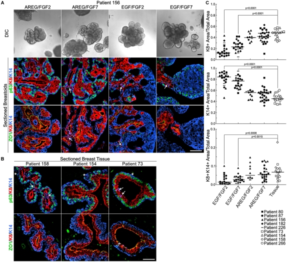
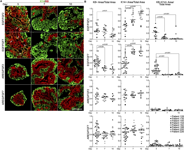
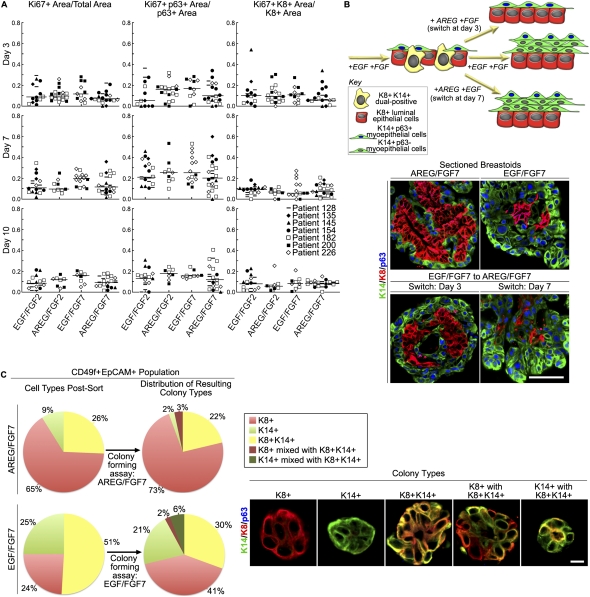
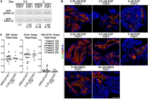
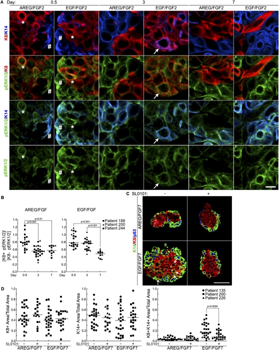
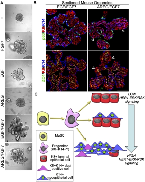
Human mammary glands arise from multipotent progenitor cells, which likely respond both to cell-autonomous and to extrinsic cues. However, the identity of these cues and how they might act remain unclear. We analyzed HER1 ligand effects on mammary morphogenesis using a three-dimensional organoid model generated from human breast tissue that recapitulates both qualitatively and quantitatively the normal ductal network in situ. Strikingly, different HER1 ligands generate distinct patterns of cell fate. Epidermal growth factor (EGF) causes a massive expansion of the myoepithelial lineage. Amphiregulin, in contrast, enables normal ductal development. These differences cannot be ascribed to preferential apoptosis or proliferation of differentiated cell populations, but are dependent on HER1 signal intensity. Inhibition of the extracellular signal-regulated kinase 1/2 (ERK1/2) effector RSK prevents the EGF-induced myoepithelial expansion. Notably, mouse mammary organoids are much less responsive to HER1 ligands. Little is known about the myoepithelial lineage or about growth factor effects on mammary progenitor differentiation, and our studies provide an important window into human mammary development that reveals unexpected differences from the mouse model.


Figures

Figure 1.
Figure 1.
AREG and an FGFR2 ligand recapitulate the ductal structure observed in situ in the human breast. (A) Representative DIC images and sections of immunostained paraffin-embedded breastoids at 29 d after culturing in response to the indicated growth factor combinations. Immunostained for luminal epithelial (K8 [red]), myoepithelial (K14 [blue]), and p63 [green]), and the tight junction marker ZO-1 (green). Arrows indicate dual-positive K8+K14+ cells. FGF2 was used at 2 nM and all growth factors were used at 5 nM. Bars: DIC, 100 μm; sections, 50 μm. (B) Representative sections of immunostained paraffin-embedded breast tissue were obtained from the normal margin of a breast cancer (158) or from breast reduction surgeries (154 and 73). Asterisks indicate K14+p63 cells. Bar, 50 μm. (C) Scatter plot showing the fraction of different cell types that comprise the ductal structure of breast tissue or breastoids. The bar represents the median. F-test of data (Supplemental Table 2) showing that intrapatient and interpatient variability are not statistically different. Statistical significance was determined using the Student's t-test and all P-values ≤0.01 were reported.
Figure 2.
Figure 2.
EGF alters ductal morphology early in development. (A) Representative images of immunostained paraffin-embedded breastoids after culturing in various growth factor combinations for differing lengths of time. The arrows show dual-positive K8+K14+ cells. Bar, 50 μm. (B) Scatter plots showing the percentage of different cell types at the indicated times after culturing in various growth factor combinations. The bar represents the median. F-test of data (Supplemental Table 3) showing that intrapatient and interpatient variability are not statistically different. Statistical significance was determined using the one-way analysis of variance (ANOVA) and all P-values ≤0.01 were reported.
Figure 3.
Figure 3.
EGF regulates myoepithelial lineage determination. (A) Comparison of the proliferation of different cell types at various times after culturing in response to different growth factor combinations. The bar represents the median. F-test of data (Supplemental Table 4) showing that intrapatient and interpatient variability are not statistically different. Statistical significance was determined using the Student's t-test and all P-values ≤0.01 were reported. (B) Schematic showing the design of the media switch experiment. Representative sections of immunostained paraffin-embedded breastoids. In the bottom panels, the medium was switched from EGF/FGF7 to AREG/FGF7 at the third or seventh day of culturing. Breastoids were cultured until day 10. Bar, 50 μm. (C) Immunofluorescence analysis of cells isolated by FACS from breastoids cultured for 3 d in AREG/FGF7 or EGF/FGF7. Cells (5000) from the CD49F+EpCAM+ population were cultured in Matrigel and maintained in the original growth factor conditions for 7 d. The subsequent colonies were analyzed by immunofluorescence and scored by the indicated phenotypes. Bar, 10 μm. Analysis was repeated on breastoids obtained from three separate patients.
Figure 4.
Figure 4.
Ligand affinity for HER1 controls lineage determination. (A) Immunoblot of breastoids cultured for 3 d with AREG/FGF7 or EGF/FGF7. Normalized levels of pERK1/2 ± range relative to total ERK1/2 (n = 2). (B) Representative sections of paraffin-embedded breastoids treated with HER1 ligands at the indicated concentration in the presence of FGFR2 ligands. After culturing for 12 d, the breastoids were analyzed. Bar, 50 μm. (C) Scatter plot showing the percentage of different cell types after culturing in low-dose EGF (0.5 nM)/FGF7 or AREG (5.0 nM)/FGF7.
Figure 5.
Figure 5.
HER1–ERK1/2–RSK signaling regulates ductal development. (A) Representative sections of paraffin-embedded breastoids treated with varying growth factor conditions for differing lengths of time. Sections were immunostained with pERK1/2, K8, and K14. The hash mark (#), asterisk (*), and arrow show K14+, K8+, and dual-positive cells, respectively, with high levels of pERK1/2. Bar, 10 μm. (B) Scatter plot comparing the relative proportion of pERK1/2 intensity in K8+ compared with K8 cells after culturing in various growth factor conditions for differing lengths of time. The bar represents the median. Data using FGF2 or FGF7 were pooled. F-test of data (Supplemental Table 5) showing that intrapatient and interpatient variability are not statistically different. Statistical significance was determined using the one-way ANOVA and all P-values ≤0.01 were reported. (C) Representative images of immunostained paraffin-embedded breastoids treated with AREG or EGF with FGF7 in the presence or absence of 100 μM SL0101 for 3 d. The arrow shows dual-positive K8+K14+ cells. Bar, 50 μm. (D) Scatter plot showing the percentage of different cell types after culturing in various growth factor combinations in the absence or presence of SL0101 for 3 d. The bar represents the median. F-test of data (Supplemental Table 6) showing that intrapatient and interpatient variability are not statistically different. Statistical significance was determined using the Student's t-test and all P-values ≤0.01 were reported.
Figure 6.
Figure 6.
Mouse mammary organoids generate normal ducts in response to EGF/FGF7. (A) Representative DIC images of mouse mammary organoids at day 3 after culturing with the indicated growth factors. Bar, 100 μm. (B) Representative sections of immunostained paraffin-embedded mouse mammary organoids that were cultured for 7 d. Arrowheads indicate K8+ cells that are not surrounded by K14+ cells. Bar, 50 μm. (C) Schematic showing the effect of HER1 signaling intensity on lineage determination in human ductal development.

References

    1. Aguirre A, Rubio ME, Gallo V 2010. Notch and EGFR pathway interaction regulates neural stem cell number and self-renewal. Nature 467: 323–327 - PMC - PubMed
    1. Andrechek ER, White D, Muller WJ 2005. Targeted disruption of ErbB2/Neu in the mammary epithelium results in impaired ductal outgrowth. Oncogene 24: 932–937 - PubMed
    1. Asselin-Labat ML, Vaillant F, Sheridan JM, Pal B, Wu D, Simpson ER, Yasuda H, Smyth GK, Martin TJ, Lindeman GJ, et al. 2010. Control of mammary stem cell function by steroid hormone signalling. Nature 465: 798–802 - PubMed
    1. Baldys A, Gooz M, Morinelli TA, Lee MH, Raymond JR Jr, Luttrell LM, Raymond JR Sr 2009. Essential role of c-Cbl in amphiregulin-induced recycling and signaling of the endogenous epidermal growth factor receptor. Biochemistry 48: 1462–1473 - PMC - PubMed
    1. Barsky SH, Karlin NJ 2006. Mechanisms of disease: breast tumor pathogenesis and the role of the myoepithelial cell. Nat Clin Pract Oncol 3: 138–151 - PubMed

Publication types

MeSH terms