Review
. 2011 Aug;188(4):773-82.
doi: 10.1534/genetics.111.131433.

Genome engineering with zinc-finger nucleases

Affiliations
Review

Genome engineering with zinc-finger nucleases

Dana Carroll. Genetics. 2011 Aug.

Abstract

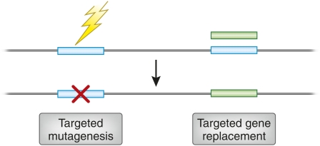
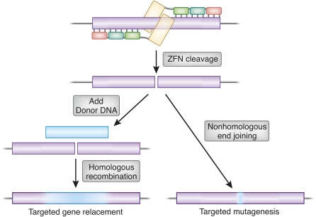
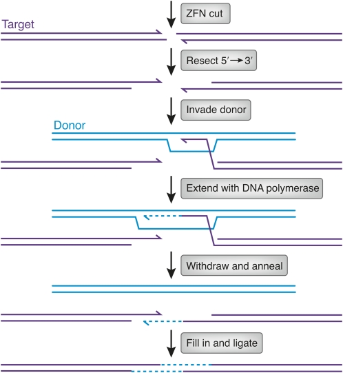
Zinc-finger nucleases (ZFNs) are targetable DNA cleavage reagents that have been adopted as gene-targeting tools. ZFN-induced double-strand breaks are subject to cellular DNA repair processes that lead to both targeted mutagenesis and targeted gene replacement at remarkably high frequencies. This article briefly reviews the history of ZFN development and summarizes applications that have been made to genome editing in many different organisms and situations. Considerable progress has been made in methods for deriving zinc-finger sets for new genomic targets, but approaches to design and selection are still being perfected. An issue that needs more attention is the extent to which available mechanisms of double-strand break repair limit the scope and utility of ZFN-initiated events. The bright prospects for future applications of ZFNs, including human gene therapy, are discussed.


Figures

Figure 1
Figure 1
Illustration of two types of genome engineering. In the top portion, the horizontal line represents a genome segment, and the open rectangles, two individual genes. The jagged arrow on the left indicates an unspecified mutagenic agent targeted to one gene. The shaded rectangle on the right is a manipulated version of the second gene that has been supplied by the experimenter. The outcomes below are targeted mutagenesis, resulting in a localized sequence alteration (“x”), and targeted gene replacement, produced by homologous recombination between the original and exogenous gene copies.
Figure 2
Figure 2
Repair outcomes of a genomic double-strand break, illustrated for the case of ZFN cleavage. A pair of three-finger ZFNs is shown at the top in association with a target gene (open box). If a homologous donor DNA is provided (solid box, left), repair can proceed by homologous recombination using the donor as template. The amount of donor sequence ultimately incorporated will typically decline with distance from the original break, as illustrated by the shading. Alternatively, the break can be repaired by nonhomologous end joining, leading to mutations at the cleavage site. These may be deletions, insertions, and base substitutions, usually quite localized, but sometimes extending away from the break.
Figure 3
Figure 3
Illustration of a pair of ZFNs bound to DNA. Zinc fingers are shown as open boxes, with short vertical lines indicating the main contacts with the DNA base pairs. FokI cleavage domains are shown as shaded boxes, with common cleavage sites, spaced by 4 bp, and indicated by vertical arrows. Zinc fingers are numbered from the N terminus. The linker between the binding and cleavage domains of one protein is labeled. The spacer between the zinc-finger binding sites, 6 bp in this case, is also indicated.
Figure 4
Figure 4
Model of a pair of ZFNs bound to DNA. Each zinc finger is shown in a shade of pink, in ribbon representation on the left and space-filling representation on the right. The FokI cleavage domains are shown in shades of blue. The four-amino-acid linker between the binding and cleavage domains is gray. DNA is shown with the sugar–phosphate backbone in orange and the bases in orange and blue. The separation between ZF binding sites is 6 bp. This model (Smith et al. 2000) was compiled from crystal structures of zinc fingers bound to DNA (Protein Database 1MEY) and the FokI restriction endonuclease in the absence of DNA (2FOK). I am grateful to Dr. Frank Whitby for help with the modeling.
Figure 5
Figure 5
Illustration of the synthesis-dependent strand annealing mechanism of homologous recombination. After ZFN cleavage, the ends of the target DNA are resected by 5′ → 3′ exonuclease action (3′ ends are shown with half arrowheads). One of the resulting single-stranded 3′ ends invades homologous sequences in the donor (thick lines). The invading 3′ end is extended by DNA polymerase (dashed line). After some synthesis, the extended end withdraws and anneals to the other end at the original break. The gaps are filled in (dashed lines; thick lines denote donor sequence, thin lines target sequence), and continuity of the strands is restored by ligation. The extent of donor sequence incorporated at the target depends on (1) the extent of synthesis after invasion, (2) whether the invading 3′ end had been chewed back, and (3) the direction of mismatch repair in the heteroduplex formed by annealing.

References

    1. Alwin S., Gere M. B., Gulh E., Effertz K., Barbas C. F., III, et al. , 2005. Custom zinc-finger nucleases for use in human cells. Mol. Ther. 12: 610–617 - PubMed
    1. Ashworth J., Havranek J. J., Duarte C. M., Sussman D., Monnat R. J., Jr, et al. , 2006. Computational redesign of endonuclease DNA binding and cleavage specificity. Nature 441: 656–659 - PMC - PubMed
    1. Beumer K., Bhattacharyya G., Bibikova M., Trautman J. K., Carroll D., 2006. Efficient gene targeting in Drosophila with zinc finger nucleases. Genetics 172: 2391–2403 - PMC - PubMed
    1. Beumer K. J., Trautman J. K., Bozas A., Liu J.-L., Rutter J., et al. , 2008. Efficient gene targeting in Drosophila by direct embryo injection with zinc-finger nucleases. Proc. Natl. Acad. Sci. USA 105: 19821–19826 - PMC - PubMed
    1. Bibikova M., Carroll D., Segal D. J., Trautman J. K., Smith J., et al. , 2001. Stimulation of homologous recombination through targeted cleavage by chimeric nucleases. Mol. Cell. Biol. 21: 289–297 - PMC - PubMed

Publication types