. 2011 Aug 9;30(18):3776-85.
doi: 10.1038/emboj.2011.271.

The structure of a transcription activation subcomplex reveals how σ(70) is recruited to PhoB promoters

Affiliations

The structure of a transcription activation subcomplex reveals how σ(70) is recruited to PhoB promoters

Alexandre G Blanco et al. EMBO J. .

Abstract

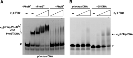
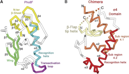
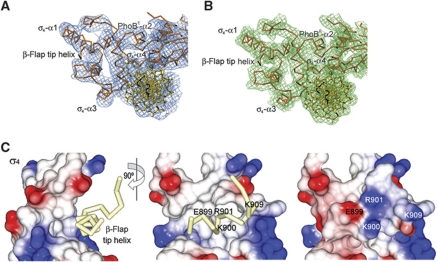
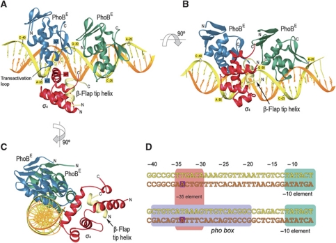
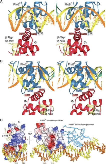
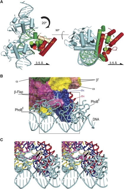
PhoB is a two-component response regulator that activates transcription by interacting with the σ(70) subunit of the E. coli RNA polymerase in promoters in which the -35 σ(70)-recognition element is replaced by the pho box. The crystal structure of a transcription initiation subcomplex that includes the σ(4) domain of σ(70) fused with the RNA polymerase β subunit flap tip helix, the PhoB effector domain and the pho box DNA reveals how σ(4) recognizes the upstream pho box repeat. As with the -35 element, σ(4) achieves this recognition through the N-terminal portion of its DNA recognition helix, but contact with the DNA major groove is less extensive. Unexpectedly, the same recognition helix contacts the transactivation loop and helices α2 and α3 of PhoB. This result shows a simple and elegant mechanism for polymerase recruitment to pho box promoters in which the lost -35 element contacts are compensated by new ones with the activator. In addition, σ(4) is reoriented, thereby suggesting a remodelling mechanism for transcription initiation.


Conflict of interest statement

The authors declare that they have no conflict of interest.

Figures

Figure 1
Figure 1
σ4-β-Flap tip helix chimera binding to DNA. EMSA of the σ4-β-flap tip helix/PhoBE/pho box DNA ternary complex. (A) Increasing amounts of σ4-β-flap tip helix chimera (0, 0.055, 0.11 and 0.18 nmol, respectively) were added to a pre-formed PhoBE/pho box DNA complex (lanes 2–5) or to a free pho box (lanes 6–8). The positions of the PhoBE/pho box DNA complex and the σ4-β-flap/PhoBE/pho box DNA are indicated (arrow and bracket, respectively). F, free DNA probe. (B) σ4-β-Flap tip helix interaction with a −35 DNA probe. Increasing amounts of σ4-β-flap tip helix (0, 0.055, 0.11 and 0.18 nmol, respectively) were added either to pho box DNA (lanes 1–4) or to a DNA probe containing the canonical −35 motif (see Materials and methods). The position of the σ4-β-flap tip helix/−35 DNA probe is indicated (arrow). F, free DNA probe.
Figure 2
Figure 2
Overview of protein–DNA structures. Cα traces of transcriptional activator PhoBE (A) and σ4-β-flap tip helix chimera (B) highlighting important structural elements in different colours.
Figure 3
Figure 3
Electron density maps of the σ4-β-flap/PhoBE/pho box DNA complex and details of the σ4-β-flap tip helix chimera. (A) Experimental σA-weighted electron density map of the ternary complex at 6.5 Å after density modification (see Materials and methods), contoured at 1σ. The final model is shown fitted in the density. (B) Final refined σA-weighted electron density map of the ternary complex at 4.3 Å. (C) Electrostatic potential surface representation of σ4 showing the β-flap tip helix as a Cα trace in pale yellow (left and middle panels) in two views 90° apart. The β-flap tip helix fits in a hydrophobic crevice of the σ4 surface. At the right panel (same view as the middle panel), the electrostatic potential surface representation includes the attached β-flap tip helix.
Figure 4
Figure 4
Structure of the σ4-β-flap tip helix chimera/PhoBE/pho box DNA complex. (A) Ribbon diagrams of the ternary complex. The upstream and the downstream protomers of PhoBE are shown in blue and green, respectively; the coding and template DNA strands are coloured in yellow and orange, respectively; the chimera is shown as σ4 in red and the β-flap tip helix in pale yellow. Note that the σ4 subunit interacts only with the upstream PhoBE. (B, C) Different orientations of the ribbon representation in (B) to show the relative positioning of the different multisubunits of the ternary complex. (D) Alignment of an RNAPH promoter containing the canonical −35 and −10 elements (upper sequence) and the phoA promoter containing the pho box (lower sequence). In the ternary complex reported here, the PhoBE dimer covers the whole pho box sequence (light violet rectangle), while σ4 covers the sequence from −35 to −30 (pink rectangle) which coincides with the binding site of σ4 onto the canonical −35 element (upper sequence, pink rectangle). Two conserved adenines at position −34 in both sequences are highlighted in dark violate squares (see text).
Figure 5
Figure 5
Interactions within the ternary complex. (A) Top: ribbon stereo plot showing the interactions between σ4 (red) and the pho box DNA. Interacting amino acids and DNA bases are indicated. (B) Stereo plot showing the interactions of σ4 (red) with the PhoBE upstream protomer (blue). The view rotated 180° about the horizontal axis relative to (A). (C) Electrostatic potential surface representation of the σ4 domain bound to the DNA (left) or to the upstream protomer of the PhoBE tandem (right) within the ternary complex. The σ4/PhoBE complex has been ‘opened’ by swinging the σ4 domain 180° to show the electrostatic potential of the interaction surface. The residues from each subunit that confront the residues from the other subunit in a contacting distance have been indicated. A base from each DNA strand is also shown to make the 180° turn of the structure clearer. The β-flap tip helix (pale yellow) and the downstream PhoBE protomer (light blue) are shown as Cα traces.
Figure 6
Figure 6
Displacement of σ4 upon binding to the pho box. (A) Two views of the superposition of the ternary complex reported here and the complex σ4-DNA (1KU7; Campbell et al, 2002); superposition was done by matching the DNA phosphates from the pho box (our structure) and the −35 element (1KU7). Schematic representations of the σ4 domains of σ70 and σA are depicted in red and green, the β-flap tip helix in cream and the PhoBE dimer in cyan. The σ4 domain of σ70 is partly displaced from the DNA as a result of the presence of PhoBE. (B) Fitting of the PhoB-DNA and σ4-β-flap tip helix chimera structure against the RNAPH structure. Colour codes for σ4, the β-flap tip helix and the PhoBE dimer are as in (A) for our ternary complex. The σ4 domain as it is in the RNAPH is shown in blue. RNAPH subunits are differentiated by colours and labelled. The superposition shows that the upstream PhoBE protomer contacts and reorientates the σ4 domain. (C) Close-up stereo view of the area indicated as a square in (B). The σ4 movement would drag the β-flap tip helix, thus facilitating the exit of nascent RNA. The black arrow indicates the movement of the β-flap tip helix, from the position observed in the RNAPH structure (β-flap tip helix in lilac) to the position observed in the present structure, when PhoBE is bound at the Pho promoter.

Similar articles

Cited by

References

    1. Abrahams JP, Leslie AG (1996) Methods used in the structure determination of bovine mitochondrial F1 ATPase. Acta Crystallogr D Biol Crystallogr 52: 30–42 - PubMed
    1. Benoff B, Yang H, Lawson CL, Parkinson G, Liu J, Blatter E, Ebright YW, Berman HM, Ebright RH (2002) Structural basis of transcription activation: the CAP-alpha CTD-DNA complex. Science 297: 1562–1566 - PubMed
    1. Blanco AG, Solà M, Gomis-Rüth FX, Coll M (2002) Tandem DNA recognition by PhoB, a two-component signal transduction transcriptional activator. Structure 10: 701–713 - PubMed
    1. Brünger AT, Adams PD, Clore GM, DeLano WL, Gros P, Grosse-Kunstleve RW, Jiang J-S, Kuszewski J, Nilges M, Pannu NS, Read RJ, Rice LM, Simonson T, Warren GL (1998) Crystallography & NMR System: a new software suite for macromolecular structure determination. Acta Crystallogr D Biol Crystallogr 54: 905–921 - PubMed
    1. Burgess RR (1969) Separation and characterization of the subunits of ribonucleic acid polymerase. J Biol Chem 244: 6168–6176 - PubMed

Publication types

MeSH terms