

doi: 10.1371/journal.pbio.1001120.
Epub 2011 Aug 2.
A case of convergence: why did a simple alternative to canonical antibodies arise in sharks and camels?
Affiliations
- PMID: 21829328
- PMCID: PMC3149040
- DOI: 10.1371/journal.pbio.1001120
Item in Clipboard
A case of convergence: why did a simple alternative to canonical antibodies arise in sharks and camels?
PLoS Biol.
2011 Aug.
No abstract available
Conflict of interest statement
The authors have declared that no competing interests exist.
Figures
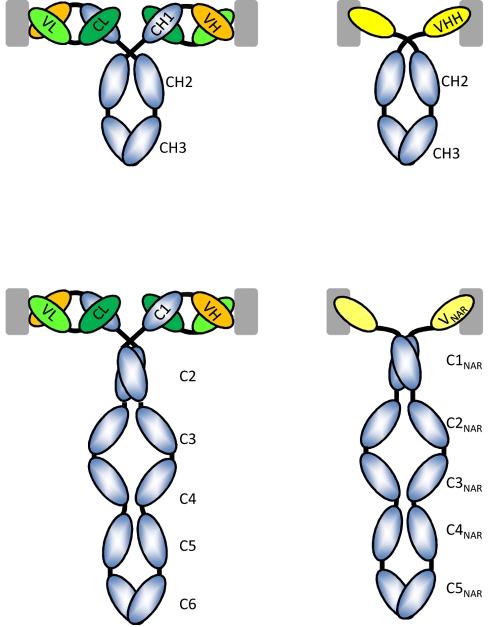
The left column displays the classical antibodies with two identical H-chains and two identical L-chains as they occur in mammals (IgG, top) and cartilaginous fish (IgW, bottom). The L-chain is in green and the antigen-binding site is formed by the paired VH and VL domains. The top right figure is the HCAb as it occurs in sera of camelids, the CH1 domain is missing and there is no L-chain. The antigen-binding site consists of one single domain, known as VHH. The H-chain of the IgW comprises six C domains and a variable domain at the N-terminal end, whereas the IgNAR (bottom right) is a homodimer of a H-chain with five C domains and a V-NAR at its N-terminal end. Note that the equivalent of the first C domain is absent. All of the antibodies are bivalent and the recognition of a possible antigen (gray square) is shown. The VH-VL associated preferentially with flat surfaces on the antigen, whereas the VHH or V-NAR has a preference to interact with cavities on the surface of the antigen.
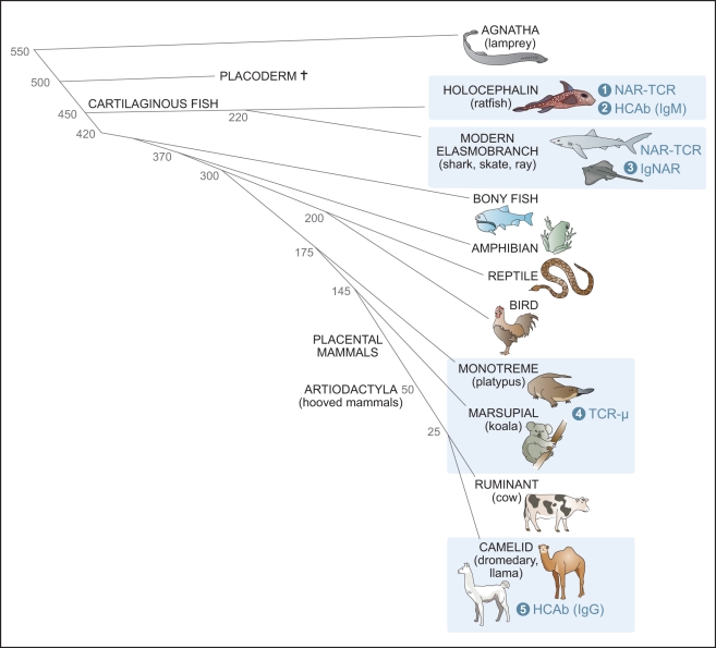
Blue boxes display animal groups shown, or predicted, to have single-domain variable regions. There are five known single domains, at least four of which were derived by convergent evolution, holocephalin HCAb, elasmobranch IgNAR, monotreme/marsupial Igμ, and camelid HCAb. The numbers on the left indicate divergence time (millions of years ago) for the various vertebrate taxa.
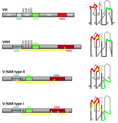
At left are displayed the linear sequence hallmarks such as the complementarity determining region (CDR) within the more conserved framework (FR) residues (gray). The disulfide bonds connecting the CDR3 with the CDR1 or FR in a VHH or V-NAR are shown by the yellow line. The folded structures of the V domains with the nine or seven β strands (named A to G in V-NARs, and A to G with insertion of strands C′ and C′′ between C and D for VH and VHH) forming two β-sheets are shown on the right. The purple diamonds on the structure denote the VHH hallmark residues in FR2 or the polar charged residues in the V-NARs. The N- and C-terminal ends of the polypeptides are shown in the structure of VHH and V-NAR type I.
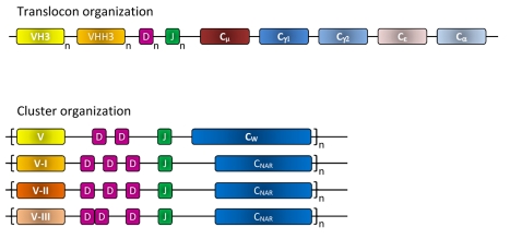
The translocon organization (top) of the gene segments as it occurs in camelids to produce the H-chain of classical antibodies and the H-chain of HCAbs. For the former antibody type, one member of the different VH families (shown VH4 and VH3) are rearranged with one of the D and one of the J genes to be transcribed with Cµ and after a class switch with the Cγ1, Cε, or Cα. Note that the greater the number of families and the more members per family, the larger the repertoire will be. For the production of HCAbs, one of the VHH3 genes (or the VH4) has to be rearranged with one of the D and one of the J genes of the same translocon before transcription occurs with the Cµ, and after a class switch, with the Cγ2 (or Cγ3 not shown). To generate the H chain of an H2L2 antibody, a VH3 gene (or VH4) has to recombine with one D and one J minigene and should be transcribed with the Cµ, or after a class switch with any other C isotype except Cγ2 or Cγ3. Cartilaginous fish have a cluster organization of their antibody genes whereby each cluster contains a dedicated V element followed by two or three D genes and a J gene. After rearrangement of these minigenes, cotranscription occurs with the W constant gene or with the C-NAR genes to produce classical H2L2 antibodies or HCAbs, respectively. There are multiple clusters in the genome of shark and the V minigenes of some clusters belong to either type I, type II (both clustered with 3-D segments), or type III (clustered with 2-D segments) minigenes.
References
-
- Hamers-Casterman C, Atarhouch T, Muyldermans S, Robinson G, Hamers C, et al. Naturally occurring antibodies devoid of light chains. Nature. 1993;363:446–448. - PubMed
-
- Greenberg A. S, Avila D, Hughes M, Hughes A, McKinney E. C, et al. A new antigen receptor gene family that undergoes rearrangement and extensive somatic diversification in sharks. Nature. 1995;374:168–173. - PubMed
-
- Muyldermans S, Cambillau C, Wyns L. Recognition of antigens by single-domain antibody fragments: the superfluous luxury of paired domains. Trends Biochem Sci. 2001;26:230–235. - PubMed
-
- Dooley H, Flajnik M. F. Antibody repertoire development in cartilaginous fish. Dev Comp Immunol. 2006;30:43–56. - PubMed
Publication types
MeSH terms
Substances
LinkOut - more resources
Full Text Sources
Other Literature Sources
