. 2011 Jul;7(7):e1002206.
doi: 10.1371/journal.pgen.1002206. Epub 2011 Jul 28.

Stress-induced PARP activation mediates recruitment of Drosophila Mi-2 to promote heat shock gene expression

Affiliations

Stress-induced PARP activation mediates recruitment of Drosophila Mi-2 to promote heat shock gene expression

Magdalena Murawska et al. PLoS Genet. 2011 Jul.

Abstract

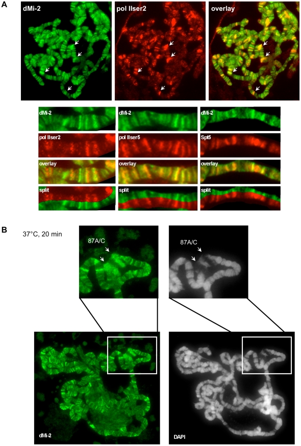
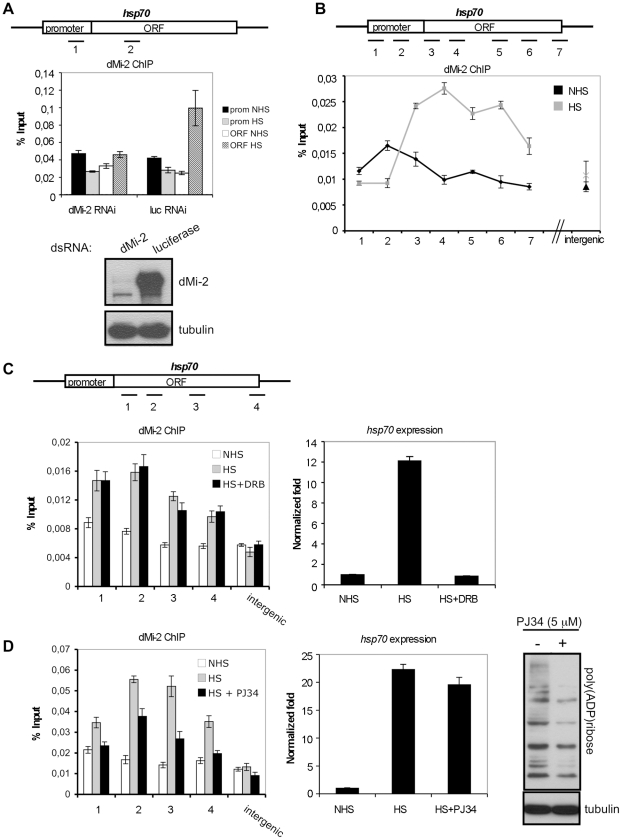
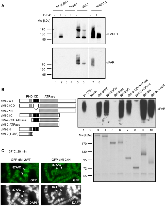
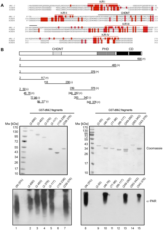
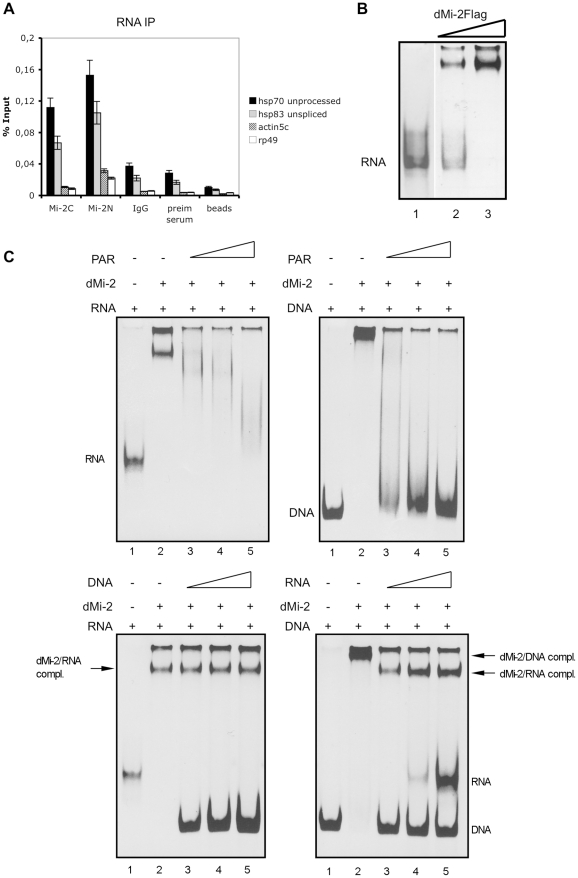
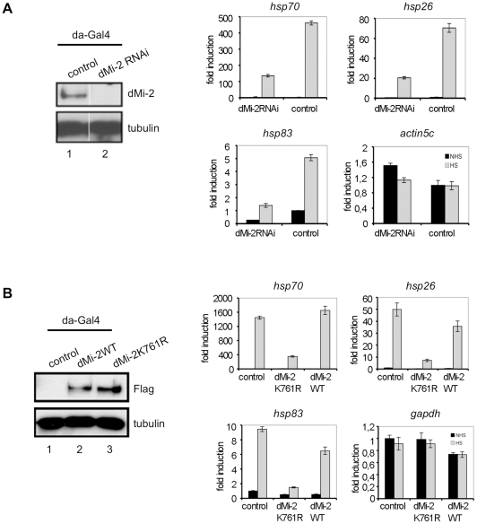
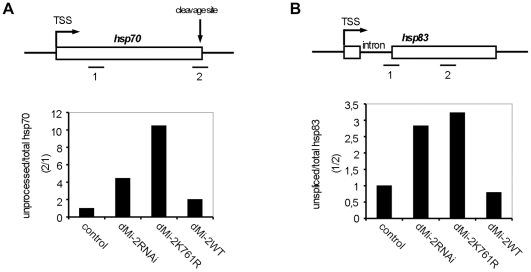
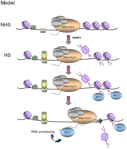
Eukaryotic cells respond to genomic and environmental stresses, such as DNA damage and heat shock (HS), with the synthesis of poly-[ADP-ribose] (PAR) at specific chromatin regions, such as DNA breaks or HS genes, by PAR polymerases (PARP). Little is known about the role of this modification during cellular stress responses. We show here that the nucleosome remodeler dMi-2 is recruited to active HS genes in a PARP-dependent manner. dMi-2 binds PAR suggesting that this physical interaction is important for recruitment. Indeed, a dMi-2 mutant unable to bind PAR does not localise to active HS loci in vivo. We have identified several dMi-2 regions which bind PAR independently in vitro, including the chromodomains and regions near the N-terminus containing motifs rich in K and R residues. Moreover, upon HS gene activation, dMi-2 associates with nascent HS gene transcripts, and its catalytic activity is required for efficient transcription and co-transcriptional RNA processing. RNA and PAR compete for dMi-2 binding in vitro, suggesting a two step process for dMi-2 association with active HS genes: initial recruitment to the locus via PAR interaction, followed by binding to nascent RNA transcripts. We suggest that stress-induced chromatin PARylation serves to rapidly attract factors that are required for an efficient and timely transcriptional response.


Conflict of interest statement

The authors have declared that no competing interests exist.

Figures

Figure 1
Figure 1. dMi-2 is recruited to HS genes.
Immunofluorescence (IF) staining of polytene chromosomes with dMi-2, RNA polymerase II (pol IIser2 and pol IIser5), Spt5 antibodies and DAPI as indicated. (A) Untreated chromosomes. Arrows show prominent sites of colocalization. Lower panels show magnified sections of individual chromosome arms. (B) Heat shocked chromosomes. Upper panels show magnified section containing the hsp70 loci 87A and 87C (arrows).
Figure 2
Figure 2. dMi-2 recruitment to HS genes requires PARP activity.
(A and B) ChIP analyses of dMi-2 binding to the hsp70 gene in Kc cells. (A) Upper panel: hsp70 gene and position of amplimers analysed (1: centred at -154, 2: +681). Middle panel: dMi-2 ChIP from cells treated with dsRNA against luciferase or dMi-2 as indicated. prom (amplimer 1): promoter; ORF (amplimer 2): open reading frame; NHS: non heat shock; HS: heat shock. Lower panel: Verification of RNAi knockdown by Western blot. (B) Upper panel: hsp70 gene and position of amplimers analysed (1: centred at -350; 2: -154; 3: +58; 4: +681; 5: +1702; 6: +2065; 7: +2549). Lower panel: dMi-2 ChIP from NHS (black graph) and HS (gray graph) cells. (C and D) Effect of elongation inhibitor DRB (C) and PARP inhibitor PJ34 (D) on dMi-2 recruitment to hsp70 gene. hsp70 gene and position of amplimers analysed are shown on top (1: centred at +58; 2: +681; 3: +1702; 4: +2549). Left panels: ChIP analyses of dMi-2 binding to hsp70 gene. Right panels: RT-QPCR analysis of hsp70 transcription. (D) Rightmost panel: anti-PAR Western blot of extracts from untreated and PJ34 treated Kc cells.
Figure 3
Figure 3. dMi-2 binds PAR.
(A) PAR was synthesised in vitro by recombinant PARP1 in the presence (+) or absence (-) of PJ34. Reactions were incubated with control anti-Flag beads (beads) and beads loaded with dMi-2 or mH2a1.1 as indicated on top. Lanes 1, 2: input; Bound material was analysed by Western blot using PARP1 (upper panel) and PAR (lower panel) antibodies. (B) Mapping PAR binding regions. Left panel: Schematic representation of dMi-2 constructs used. Amino acid boundaries are as follows: dMi-2WT: dMi-2 1-1982; dMi-2ΔCD: dMi-2 Δ485-690; dMi-2ΔN: 691-1982; dMi-2ΔC: dMi-2 1-1271; dMi-2-CD+ATPase: dMi-2 484-1271; dMi-2-ATPase: dMi-2 691-1271; dMi-2N: dMi-2 1-690; dMi-2(1-485): dMi-2 1-485. Upper right panel: PAR binding assays with dMi-2 mutants were performed as in (A). dMi-2 mutants are shown on top. Bound material was analysed by anti-PAR Western blot. Lane 1: input. Lower right panel: Coomasie stained gel showing the dMi-2 constructs used. (C) Polytene chromosomes from transgenic larvae expressing GFP-dMi-2 transgenes (dMi-2WT and dMi-2ΔN) were analysed by IF using GFP antibody (green) and DAPI (gray). Arrows point to hsp70 HS loci 87A and 87C.
Figure 4
Figure 4. PAR–binding regions of dMi-2.
(A) Multiple sequence alignment of N-terminus of dMi-2 and human and mouse CHD4. All K and R amino acid residues are coloured in red. Red lines indicate the four K/R rich regions. The black line indicates the CHDNT domain. (B) Mapping of PAR binding regions in the N-terminal part of dMi-2. Upper panel: Schematic representation of dMi-2 constructs used. Numbers indicate the amino acid borders of the constructs. (+) and (-) indicate binding to PAR. Middle panel: Coomasie stained gels with purified GST-dMi-2 fragments used for PARP pulldown assays. Lower panel: PAR binding assays with GST-dMi-2 fragments were performed as in Figure 3B. Bound material was analysed by anti-PAR Western blot. Lanes 1 and 8: inputs.
Figure 5
Figure 5. dMi-2 binds RNA.
(A) RNA immunoprecipitation (RIP) of hsp70 and hsp83 unprocessed transcripts from heat shocked Kc cells. RIP was performed with two independent dMi-2 antibodies (dMi-2C and dMi-2N), IgG, preimmune serum and protein G beads as indicated. Primer pairs that specifically amplify actin5c, rp49 and unprocessed hsp70 and hsp83 transcripts (see Figure 7) were used for RT-QPCR. (B) RNA electrophoretic mobility shift assay. Single stranded hsp70 RNA was incubated with recombinant dMi-2. Lane 2: 0.1 µg dMi-2, lane 3: 0.2 µg dMi-2, lane 1: no protein. RNA and RNA:protein complexes were resolved by electrophoresis and visualized with ethidium bromide. Position of unbound RNA probe is indicated. (C) Competition mobility shift assays. Upper left panel: Single stranded hsp70 RNA was incubated with 0.2 µg of recombinant dMi-2 in the absence or in the presence of increasing amounts of PAR polymer, as indicated. Upper right panel: hsp70 DNA was incubated with 0.2 µg of recombinant dMi-2 in the absence or in the presence of increasing amounts of PAR polymer, as indicated. The following mass ratios of RNA to PAR or DNA to PAR were used: lane 3 - 1∶1, lane 4 -1∶2, lane 5 -1∶4. Positions of unbound RNA and DNA probes are indicated. Lower left panel: Single stranded hsp70 RNA was incubated with 0.2 µg of recombinant dMi-2 in the absence or in the presence of increasing amounts of DNA, as indicated. Lower right panel: hsp70 DNA was incubated with 0.2 µg of recombinant dMi-2 in the absence or in the presence of increasing amounts of RNA, as indicated. The following weight ratios of RNA to DNA or DNA to RNA were used: line 3 - 1∶1, lane 4 -1∶2, lane 5 -1∶4. Positions of unbound RNA and DNA probes and dMi-2/DNA and dMi-2/RNA complexes are shown on the right.
Figure 6
Figure 6. dMi-2 is required for efficient HS gene transcription.
(A) Left panel: Verification of dMi-2 knockdown in control and dMi-2 RNAi larvae. Control flies and flies carrying an dMi-2 RNAi transgene under UAS control were crossed with a da-GAL4 driver strain. Extracts from third instar larvae were subjected to Western Blot. Antibodies used are indicated on the right. Right panel: RT-QPCR analysis of hsp70, hsp26, hsp83 and actin5c expression in control and dMi-2 RNAi larvae. Values are expressed relative to the value in NHS control larvae. (B) Left panel: Verification of dMi-2 transgene expression in larvae by anti-Flag Western blot. Right panel: RT-QPCR analysis of hsp70, hsp26, hsp83 and GAPDH expression in control and transgenic larvae.
Figure 7
Figure 7. dMi-2 is required for efficient RNA processing.
(A and B) Upper panels: Schematic representations of the hsp70 and hsp83 genes. RT-QPCR amplimers, hsp70 cleavage site, hsp83 intron and transcriptional start sites (TSS) are shown. (A) Lower panel: RT-QPCR from control and transgenic larvae. The ratio between 3′ unprocessed and total hsp70 RNA was determined (hsp70: amplimer 2/amplimer 1). The ratio obtained for control larvae was set to 1, other ratios were expressed relative to this. (B) Lower panel: The ratio between unspliced and total hsp83 RNA was determined (hsp83: amplimer 1/amplimer 2) and plotted as in (A).
Figure 8
Figure 8. Model.
Upon HS, PARylation of the locus creates binding sites for PAR-sensing regions of dMi-2. dMi-2 is recruited and, subsequently, interacts with nascent transcripts to support transcription and processing. GAF: GAGA Factor, HSE: HS elements.

References

    1. Petesch SJ, Lis JT. Rapid, transcription-independent loss of nucleosomes over a large chromatin domain at Hsp70 loci. Cell. 2008;134:74–84. - PMC - PubMed
    1. Winegarden NA, Wong KS, Sopta M, Westwood JT. Sodium salicylate decreases intracellular ATP, induces both heat shock factor binding and chromosomal puffing, but does not induce hsp 70 gene transcription in Drosophila. J Biol Chem. 1996;271:26971–26980. - PubMed
    1. Tulin A, Spradling A. Chromatin loosening by poly(ADP)-ribose polymerase (PARP) at Drosophila puff loci. Science. 2003;299:560–562. - PubMed
    1. Zobeck KL, Buckley MS, Zipfel WR, Lis JT. Recruitment Timing and Dynamics of Transcription Factors at the Hsp70 Loci in Living Cells. Molecular Cell. 2010;40:965–975. - PMC - PubMed
    1. Krishnakumar R, Kraus WL. The PARP side of the nucleus: molecular actions, physiological outcomes, and clinical targets. Mol Cell. 2010;39:8–24. - PMC - PubMed

Publication types

MeSH terms

LinkOut - more resources