

Long-term decrease of retinal pigment epithelium defects in large stage iv macular holes with borders mechanically joined during surgery
- PMID: 21829404
- PMCID: PMC3150968
- DOI: 10.1159/000330553
Long-term decrease of retinal pigment epithelium defects in large stage iv macular holes with borders mechanically joined during surgery
Abstract
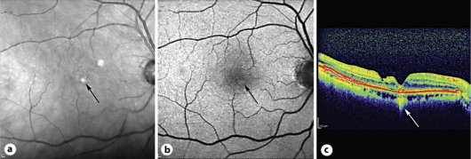
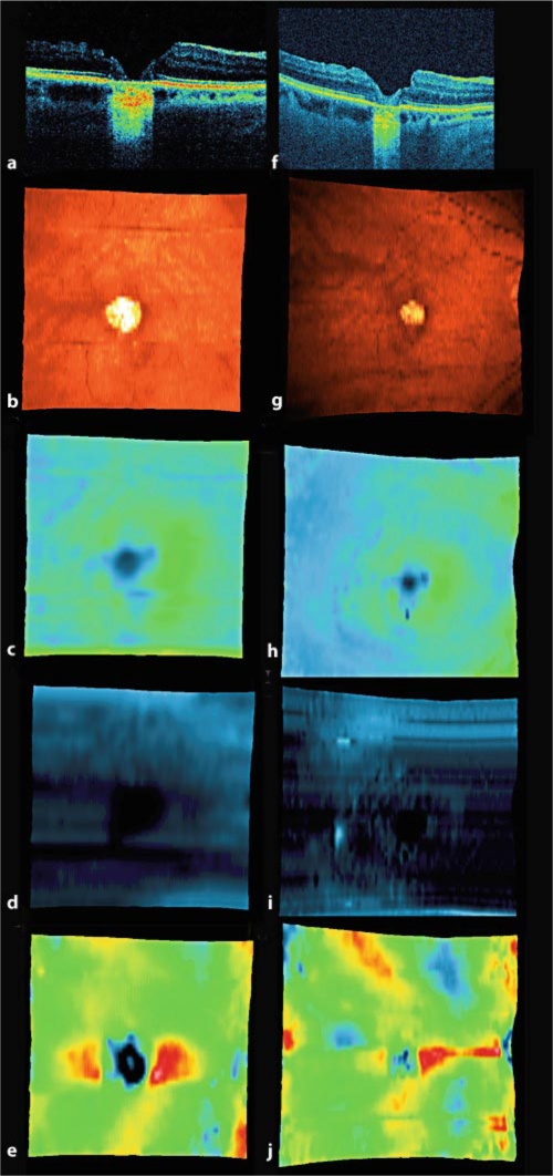
Purpose: The aim of this paper is to present retinal pigment epithelium (RPE) defects in spectral domain optical coherence tomography (SD-OCT) and their gradual resolution over time.
Materials and methods: Observational case series of 3 eyes of 3 patients who were followed for a period of 3-6 years after undergoing surgery to mechanically close the borders of large stage IV macular holes. Photoreceptor defects and RPE defects were measured during this period with SD-OCT.
Results: In all cases, a reduction in the size of the areas of photoreceptor and RPE defects was noted, which correlated with late improvement in visual acuity (VA).
Conclusions: In eyes with no underlying retinal pigment epithelial or choroidal disease, restoration of RPE is possible in vivo for up to several years after surgery for macular hole closure. An improvement in VA is possible, even lasting many years after successful macular hole surgery, which corresponds to a decrease in both RPE defects and the size of the defects in the junction between the inner and outer segments of photoreceptors.
Keywords: Macular hole; Photoreceptor defects; Retinal pigment epithelium defects; Spectral domain optical coherence tomography.
Figures
References
-
- Kelly NE, Wendel RT. Vitreous surgery for idiopathic macular holes: results of a pilot study. Arch Ophthalmol. 1991;109:654–659. - PubMed
-
- Ando F, Sasano K, Ohba N, et al. Anatomic and visual outcomes after indocyanine green-assisted peeling of the retinal internal limiting membrane in idiopathic macular hole surgery. Am J Ophthalmol. 2004;137:609–614. - PubMed
-
- Beutel J, Dahmen G, Ziegler A, Hoerauf H. Internal limiting membrane peeling with indocyanine green or trypan blue in macular hole surgery: a randomized trial. Arch Ophthalmol. 2007;125:326–332. - PubMed
-
- Alpatov S, Shchuko A, Malyshev V. A new method of treating macular holes. Eur J Ophthalmol. 2007;17:246–252. - PubMed
-
- Michalewska Z, Michalewski J, Adelman RA, Nawrocki J. Inverted internal limiting membrane (ILM) flap technique for large macular hole. Ophthalmology. 2010;117:2018–2025. - PubMed
Publication types
LinkOut - more resources
Full Text Sources
