Comparative Study
. 2011;6(7):e22485.
doi: 10.1371/journal.pone.0022485. Epub 2011 Jul 29.

Pancreatic β-cell death in response to pro-inflammatory cytokines is distinct from genuine apoptosis

Affiliations
Comparative Study

Pancreatic β-cell death in response to pro-inflammatory cytokines is distinct from genuine apoptosis

J Jason Collier et al. PLoS One. 2011.

Abstract

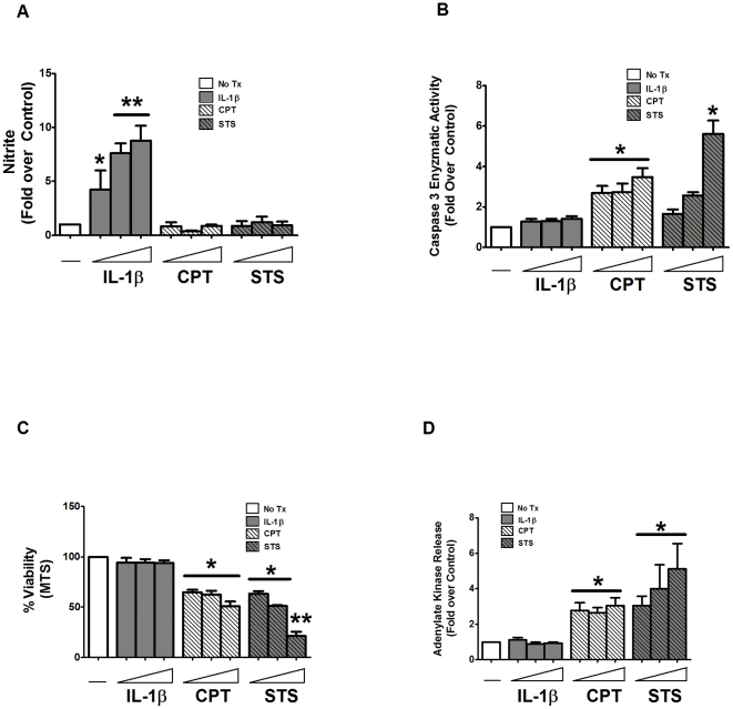
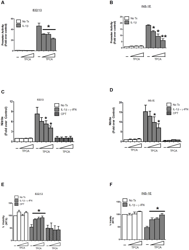
A reduction in functional β-cell mass leads to both major forms of diabetes; pro-inflammatory cytokines, such as interleukin-1beta (IL-1β) and gamma-interferon (γ-IFN), activate signaling pathways that direct pancreatic β-cell death and dysfunction. However, the molecular mechanism of β-cell death in this context is not well understood. In this report, we tested the hypothesis that individual cellular death pathways display characteristic phenotypes that allow them to be distinguished by the precise biochemical and metabolic responses that occur during stimulus-specific initiation. Using 832/13 and INS-1E rat insulinoma cells and isolated rat islets, we provide evidence that apoptosis is unlikely to be the primary pathway underlying β-cell death in response to IL-1β+γ-IFN. This conclusion was reached via the experimental results of several different interdisciplinary strategies, which included: 1) tandem mass spectrometry to delineate the metabolic differences between IL-1β+γ-IFN exposure versus apoptotic induction by camptothecin and 2) pharmacological and molecular interference with either NF-κB activity or apoptosome formation. These approaches provided clear distinctions in cell death pathways initiated by pro-inflammatory cytokines and bona fide inducers of apoptosis. Collectively, the results reported herein demonstrate that pancreatic β-cells undergo apoptosis in response to camptothecin or staurosporine, but not pro-inflammatory cytokines.


Conflict of interest statement

Competing Interests: The authors have declared that no competing interests exist.

Figures

Figure 1
Figure 1. INS-1E and 832/13 rat insulinoma cells both display sensitivity to pro-inflammatory cytokines and genuine apoptotic inducers.
832/13 and INS-1E cells were exposed to either media alone (No Tx), 1 ng/mL IL-1β+100 U/mL γ-IFN for 18 h, 2 µM camptothecin for 6 h or 1 µM staurosporine for 6 h. A. Nitrite accumulation was measured by the Griess assay. Cell viability was measured by both MTS assay (B and C), and adenylate kinase assay (D and E). Data are means ± SEM for three individual experiments. #, p = 0.05 vs. control; * p<0.05 vs. control; **, p<0.01 vs. control.
Figure 2
Figure 2. Pro-inflammatory cytokines have a dose-dependent effect on INS-1-derived cell lines.
832/13 cells were treated with standard culture media as a control (No Tx), increasing concentrations of IL-1β (0.01, 0.1, and 1 ng/mL) for 18 h, camptothecin (0.5, 1, and 2 µM) for 6 h or staurosporine (0.25, 0.5, 1 µM) for 6 h. Nitrite accumulation (A), caspase 3 enzyme activity (B), cell viability via MTS (C) or adenylate kinase release (D) were measured. Data represent means ± SEM for three individual experiments. *, p<0.05 vs. control; **, p<0.01 vs. control.
Figure 3
Figure 3. Time-dependent sensitivity of INS-1-derived cell lines in response to pro-inflammatory cytokines and bona fide apoptotic inducers.
832/13 cells were treated with media alone (No Tx), 1 ng/mL IL-1β plus 100 U/mL γ-IFN, 2 µM camptothecin or 1 µM staurosporine for 1.5, 3, and 6 h. Measurements of nitrite (A), caspase 3 enzyme activity (B), cellular viability via MTS (C) and adenylate kinase (D) were analyzed. Data are means ± SEM for three individual experiments. *, p<0.05 vs. control.
Figure 4
Figure 4. Metabolic profiling by tandem mass spectrometry reveals distinct metabolite signatures of cells exposed to pro-inflammatory cytokines vs. the apoptotic inducer camptothecin.
A. 832/13 cells were exposed to 1 ng/mL IL-1β+100 U/mL γ-IFN or 2 µM camptothecin for 6 h. Metabolite concentration was measured by mass spectrometry with values displayed as fold change relative to control. The represent values given in log2, so that “3.00” is a minimum of an 8-fold increase, while “−3.00” is a minimum of an 8-fold decrease. The red color indicates a metabolite increase in the treatment condition relative to an untreated control, while the green color represents a decrease relative to control. The p values for all metabolites are given in Tables S1 and S2. B. 832/13 cells were transduced with recombinant adenovirus expressing a luciferase reporter gene driven by three NF-κB response elements (NF-κB-luc). After 24 h, cells were cultured in increasing concentrations of camptothecin (0.5, 1, 2 µM) for 1 h followed by a 4 h incubation with 1 ng/mL IL-1β. Luciferase activity was measured using cell lysates. C–D. 832/13 and INS-1E cells were transduced with the aforementioned NF-κB-luc virus. After 24 h, these cells were then exposed to increasing concentrations of MTA (1, 2, and 4 mM) for 1 h, followed by a 4 h exposure to 1 ng/mL IL-1β. After 4 h, luciferase activity was measured. E. 832/13 cells were cultured in the presence or absence of increasing concentrations of MTA (0.5, 1, and 2 mM) for 1 h, then switched to either media alone (No Tx) or 1 ng/mL IL-1β+100 U/mL γ-IFN for 6 h, followed by measurement of nitrite accumulation in the media. *, p<0.05; **, p<0.01.
Figure 5
Figure 5. Pharmacological interference with NF-κB activity protects against cytokine-mediated cell death but not apoptosis.
A–B. 832/13 and INS-1E cells were transduced with the NF-κB-luc virus for 24 h, followed by exposure to increasing concentrations of TPCA (0.5, 1, and 2 µM) for 1 h. Luciferase activity was measured following by a subsequent 4 h culture with 1 ng/mL IL-1β. Data are means ± SE for three independent experiments, performed in duplicate each time. C–F. 832/13 and INS-1E cells were pre-treated with increasing concentrations of TPCA (0.5, 1, and 2 µM) for 1 h, followed by a 4 h treatment with either 1 ng/mL IL-1β+100 U/mL γ-IFN or 2 µM camptothecin. Nitrite was measured using the Griess assay (C and D) and viability was measured by MTS assay (E and F). Data are representative of mean ± SEM for 3–4 individual experiments. *, p<0.05; **, p<0.01.
Figure 6
Figure 6. Overexpression of the IκBα super-repressor protects against pro-inflammatory cytokines but not genuine apoptotic induction.
A. 832/13 cells were transduced with recombinant adenovirus expressing either β-galactosidase (βGal) or increasing concentrations of IκBα SR. A. Following 24 h incubation, whole cell lysates were immunoblotted for IκBα with β-actin as a loading control. Immunoblots represent 2 individual experiments. Note that HA-tagging of the IκBα SR causes it to migrate slower (top arrow) relative to the endogenous protein (bottom arrow). B. 832/13 cells were transduced with NF-κB-luc and either βGAL or the highest dose of the IκBα SR (shown in panel A) for 24 h. Cells were then treated with 1 ng/mL IL-1β for 4 h and luciferase activity was measured. Data are means ± SEM for 3 individual experiments. C–D. 832/13 cells were transduced with adenoviruses expressing either βGAL or increasing concentrations of IκBα. After 24 h, the cells were stimulated with either 1 ng/mL IL-1β or 1 µM STS for 6 h. Nitrite was measured via the Griess assay (C), and viability by MTS assay (D). Values represent means ± SEM from 4 individual experiments. *, p<0.05.
Figure 7
Figure 7. Manipulation of the apoptosome protects against apoptosis but does not impact pro-inflammatory of cytokine-mediated cell death.
A. 832/13 cells were transduced either with a recombinant adenovirus expressing βGal or dominant-negative caspase 9 for 24 h followed by a change to media alone (control) or 1 µM staurosporine for 4 h. Whole-cell lysates were immunoblotted for caspase-3 with β-actin as a loading control. Top and bottom arrows indicate total and cleaved caspase-3, respectively. Immunoblots are representative of two independent experiments. B and C. 832/13 cells were transduced with either β-Galactosidase or increasing doses of the dominant-negative caspase-9 for 24 h, followed by 4 h stimulation with either 2 µM camptothecin (B) or 1 µM staurosporine (C). Cell lysates were analyzed for caspase-3 enzyme activity and expressed as percent of the maximal response. Data are presented as mean ± SEM for three separate experiments. D. The highest dose of Caspase-9 DN from (B and C) was then used to transduced 832/13 cells. 24 h post-transduction, cells were exposed to 1 µM staurosporine for 4 h, and then phase contrast images were acquired. E. Rat islets were transduced with the indicated adenoviruses; 72 h after adenoviral transduction, the islets were exposed to 10 ng/mL IL-1β or 1 µM staurosporine for 6 h and harvested for caspase-3 enzyme activity. F-K. 832/13 cells were transduced with recombinant adenoviruses expressing either βGAL or Caspase-9 DN. After 8 h of incubation with the indicated adenoviruses, cells were then transfected with siRNA duplexes representing either a scrambled control (siScramble) or specifically targeting the APAF-1 mRNA. 24 h after siRNA transfection, cells were treated with either 2 µM camptothecin (F and H) or 1 µM staurosporine (G and I) for 4 h or 1 ng/mL IL-1β+100 U/mL γ-IFN for 18 h (J and K). *, p<0.05; **, p<0.01.
Figure 8
Figure 8. Cell death pathways activated in pancreatic β-cells in response to pro-inflammatory or apoptotic stimuli.
The combination of IL-1β+γ-IFN generates massive quantities of nitric oxide within the pancreatic β-cell which impairs β-cell function, decreases intracellular ATP levels, and inhibits apoptosome formation and/or activity, leading to cellular death by necrosis. Apoptotic signals, such as camptothecin, lead to DNA damage without nitric oxide accumulation, which signals apoptosome formation. Nitric oxide inhibits apoptosome activity but also damages DNA, which the cell attempts to repair. If the damaged DNA cannot be repaired and nitric oxide levels decrease sufficiently to allow apoptosome formation, a subset of β-cells may undergo apoptosis provided adequate ATP levels are present.

Similar articles

Cited by

References

    1. Mathis D, Vence L, Benoist C. beta-Cell death during progression to diabetes. Nature. 2001;414:792–798. - PubMed
    1. Mandrup-Poulsen T. The role of interleukin-1 in the pathogenesis of IDDM. Diabetologia. 1996;39:1005–1029. - PubMed
    1. Arnush M, Heitmeier MR, Scarim AL, Marino MH, Manning PT, et al. IL-1 produced and released endogenously within human islets inhibits beta cell function. J Clin Invest. 1998;102:516–526. - PMC - PubMed
    1. Corbett JA, Sweetland MA, Wang JL, Lancaster JR, Jr, McDaniel ML. Nitric oxide mediates cytokine-induced inhibition of insulin secretion by human islets of Langerhans. Proc Natl Acad Sci U S A. 1993;90:1731–1735. - PMC - PubMed
    1. Corbett JA, Wang JL, Sweetland MA, Lancaster JR, Jr, McDaniel ML. Interleukin 1 beta induces the formation of nitric oxide by beta-cells purified from rodent islets of Langerhans. Evidence for the beta-cell as a source and site of action of nitric oxide. J Clin Invest. 1992;90:2384–2391. - PMC - PubMed

Publication types

MeSH terms