

A single amino acid of human immunodeficiency virus type 2 capsid protein affects conformation of two external loops and viral sensitivity to TRIM5α
- PMID: 21829511
- PMCID: PMC3145752
- DOI: 10.1371/journal.pone.0022779
A single amino acid of human immunodeficiency virus type 2 capsid protein affects conformation of two external loops and viral sensitivity to TRIM5α
Abstract
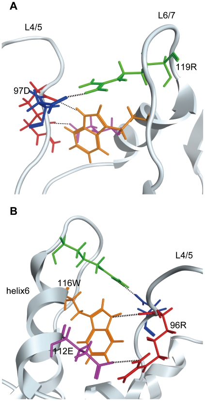
We previously reported that human immunodeficiency virus type 2 (HIV-2) carrying alanine or glutamine but not proline at position 120 of the capsid protein (CA) could grow in the presence of anti-viral factor TRIM5α of cynomolgus monkey (CM). To elucidate details of the interaction between the CA and TRIM5α, we generated mutant HIV-2 viruses, each carrying one of the remaining 17 possible amino acid residues, and examined their sensitivity to CM TRIM5α-mediated restriction. Results showed that hydrophobic residues or those with ring structures were associated with sensitivity, while those with small side chains or amide groups conferred resistance. Molecular dynamics simulation study revealed a structural basis for the differential TRIM5α sensitivities. The mutations at position 120 in the loop between helices 6 and 7 (L6/7) affected conformation of the neighboring loop between helices 4 and 5 (L4/5), and sensitive viruses had a common L4/5 conformation. In addition, the common L4/5 structures of the sensitive viruses were associated with a decreased probability of hydrogen bond formation between the 97th aspartic acid in L4/5 and the 119th arginine in L6/7. When we introduced aspartic acid-to-alanine substitution at position 97 (D97A) of the resistant virus carrying glutamine at position 120 to disrupt hydrogen bond formation, the resultant virus became moderately sensitive. Interestingly, the virus carrying glutamic acid at position 120 showed resistance, while its predicted L4/5 conformation was similar to those of sensitive viruses. The D97A substitution failed to alter the resistance of this particular virus, indicating that the 120th amino acid residue itself is also involved in sensitivity regardless of the L4/5 conformation. These results suggested that a hydrogen bond between the L4/5 and L6/7 modulates the overall structure of the exposed surface of the CA, but the amino acid residue at position 120 is also directly involved in CM TRIM5α recognition.
Conflict of interest statement
Figures


Similar articles
-
Novel mutant human immunodeficiency virus type 1 strains with high degree of resistance to cynomolgus macaque TRIMCyp generated by random mutagenesis.J Gen Virol. 2016 Apr;97(4):963-976. doi: 10.1099/jgv.0.000408. Epub 2016 Jan 20. J Gen Virol. 2016. PMID: 26795727 Free PMC article.
-
Multiple sites in the N-terminal half of simian immunodeficiency virus capsid protein contribute to evasion from rhesus monkey TRIM5α-mediated restriction.Retrovirology. 2010 Sep 8;7:72. doi: 10.1186/1742-4690-7-72. Retrovirology. 2010. PMID: 20825647 Free PMC article.
-
Modification of a loop sequence between alpha-helices 6 and 7 of virus capsid (CA) protein in a human immunodeficiency virus type 1 (HIV-1) derivative that has simian immunodeficiency virus (SIVmac239) vif and CA alpha-helices 4 and 5 loop improves replication in cynomolgus monkey cells.Retrovirology. 2009 Aug 3;6:70. doi: 10.1186/1742-4690-6-70. Retrovirology. 2009. PMID: 19650891 Free PMC article.
-
Anti-retroviral activity of TRIM5 alpha.Rev Med Virol. 2010 Mar;20(2):77-92. doi: 10.1002/rmv.637. Rev Med Virol. 2010. PMID: 20049904 Review.
-
Impact of TRIM5α in vivo.AIDS. 2015 Sep 10;29(14):1733-43. doi: 10.1097/QAD.0000000000000812. AIDS. 2015. PMID: 26372380 Free PMC article. Review.
Cited by
-
Novel mutant human immunodeficiency virus type 1 strains with high degree of resistance to cynomolgus macaque TRIMCyp generated by random mutagenesis.J Gen Virol. 2016 Apr;97(4):963-976. doi: 10.1099/jgv.0.000408. Epub 2016 Jan 20. J Gen Virol. 2016. PMID: 26795727 Free PMC article.
-
Structural insight into HIV-1 capsid recognition by rhesus TRIM5α.Proc Natl Acad Sci U S A. 2012 Nov 6;109(45):18372-7. doi: 10.1073/pnas.1210903109. Epub 2012 Oct 22. Proc Natl Acad Sci U S A. 2012. PMID: 23091002 Free PMC article.
-
A comparison of murine leukemia viruses that escape from human and rhesus macaque TRIM5αs.J Virol. 2013 Jun;87(11):6455-68. doi: 10.1128/JVI.03425-12. Epub 2013 Mar 27. J Virol. 2013. PMID: 23536686 Free PMC article.
-
Molecular dynamics simulation in virus research.Front Microbiol. 2012 Jul 19;3:258. doi: 10.3389/fmicb.2012.00258. eCollection 2012. Front Microbiol. 2012. PMID: 22833741 Free PMC article.
-
Structural dynamics of HIV-1 envelope Gp120 outer domain with V3 loop.PLoS One. 2012;7(5):e37530. doi: 10.1371/journal.pone.0037530. Epub 2012 May 18. PLoS One. 2012. PMID: 22624045 Free PMC article.
References
-
- Stremlau M, Owens CM, Perron MJ, Kiessling M, Autissier P, et al. The cytoplasmic body component TRIM5alpha restricts HIV-1 infection in Old World monkeys. Nature. 2004;427:848–853. - PubMed
Publication types
MeSH terms
Substances
LinkOut - more resources
Full Text Sources
Other Literature Sources
