. 2011;6(7):e22820.
doi: 10.1371/journal.pone.0022820. Epub 2011 Jul 28.

The regenerative capacity of the zebrafish caudal fin is not affected by repeated amputations

Affiliations

The regenerative capacity of the zebrafish caudal fin is not affected by repeated amputations

Ana Sofia Azevedo et al. PLoS One. 2011.

Abstract

Background: The zebrafish has the capacity to regenerate many tissues and organs. The caudal fin is one of the most convenient tissues to approach experimentally due to its accessibility, simple structure and fast regeneration. In this work we investigate how the regenerative capacity is affected by recurrent fin amputations and by experimental manipulations that block regeneration.

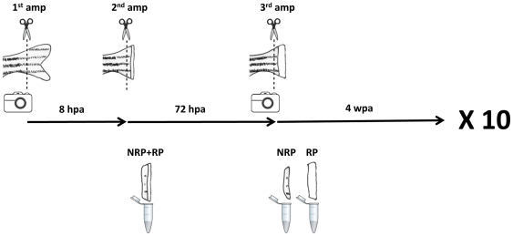
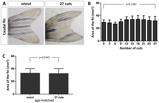
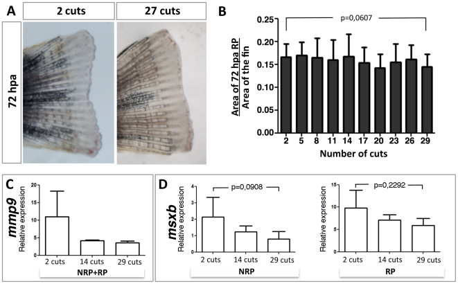
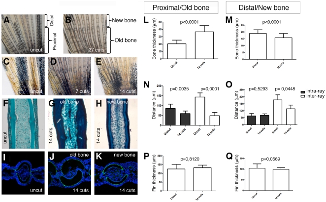
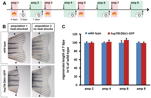
Methodology/principal findings: We show that consecutive repeated amputations of zebrafish caudal fin do not reduce its regeneration capacity and do not compromise any of the successive regeneration steps: wound healing, blastema formation and regenerative outgrowth. Interfering with Wnt/ß-catenin signalling using heat-shock-mediated overexpression of Dickkopf1 completely blocks fin regeneration. Notably, if these fins were re-amputated at the non-inhibitory temperature, the regenerated caudal fin reached the original length, even after several rounds of consecutive Wnt/ß-catenin signalling inhibition and re-amputation.

Conclusions/significance: We show that the caudal fin has an almost unlimited capacity to regenerate. Even after inhibition of regeneration caused by the loss of Wnt/ß-catenin signalling, a new amputation resets the regeneration capacity within the caudal fin, suggesting that blastema formation does not depend on a pool of stem/progenitor cells that require Wnt/ß-catenin signalling for their survival.


Conflict of interest statement

Competing Interests: The authors have declared that no competing interests exist.

Figures

Figure 1
Figure 1. Outline of the consecutive repeated caudal fin amputations performed every month over an 11-month period.
Each month, the fully regenerated caudal fin was photographed and amputated. After 8 hpa, it was subjected to a second amputation and the amputated tissue was collected. After 72 hpa, the caudal fin was photographed again, a third amputation was performed and the amputated tissues were collected. After 4 wpa, the procedure was repeated. The entire procedure was done 10 times. AMP: amputation; NRP: non-regenerate portion; RP: regenerate portion.
Figure 2
Figure 2. Consecutive repeated amputations maintain the original size of the fully regenerated caudal fin.
(A) The same caudal fin before any amputation (0 cuts) and 4 wpa after 27 consecutive cuts. (B) Area of the 4 wpa regenerated caudal fin with increasing number of cuts. (C) Comparison of the caudal fin area of zebrafish siblings that were amputated 27 consecutive times with age matched siblings that were never amputated.
Figure 3
Figure 3. The 72 hpa regenerate size of the caudal fin is maintained with consecutive repeated amputations over an 11-month period.
(A) A 72 hpa caudal fin obtained after the second consecutive amputation and after the twenty-seventh consecutive amputation. (B) Area of the 72 hpa regenerate over the area of the fully regenerated caudal fin immediately before the amputation measured with increasing number of cuts. (C) mmp9 expression levels at 8 hpa with increasing number of cuts. (D) msxb expression levels at 72 hpa in both non-regenerate portions (NRP) and regenerate portions (RP) with increasing number of cuts.
Figure 4
Figure 4. Consecutive repeated amputations affect the structure of non-regenerate bone.
Picture of the dorsal lobe of an uncut caudal fin (A) and its age-matched sibling after 27 cuts (B). Picture of the dorsal lobe of an uncut caudal fin (C) and a caudal fin after 7 (D) and 14 cuts (E). Masson's trichrome staining of longitudinal sections of an uncut bony ray (F) and of an old (G) and regenerated (H) regions of a bony ray after 14 cuts. Confocal images of transverse sections of a Zns5 immunostained proximal region of an uncut caudal fin (I) and of the old (J) and new (K) tissue of a caudal fin after 14 cuts. Quantification of the bone thickness, inter- and intra-ray tissue and fin thickness in the old (L, N, P) and new (M, O, Q).
Figure 5
Figure 5. Repeated inhibition of fin regeneration by interference with Wnt/b-catenin signaling does not diminish regenerative capacity.
(A) Schematic illustration of the experimental scheme. Red shaded areas indicate periods in which fish were heat-shocked twice daily, green areas indicate periods in which fish were allowed to regenerate in the absence of heat-shock. amp = amputation, phot = photo of the tail fin. (B) Wild-type and hsp70l:Dkk1-GFP transgenic tail fins heat-shocked until 4 dpa and photographed 7 days after amputation 1 (left column) and photographed after amputation 2 without heat-shocks (right column). Note that heat-shocked wild-type fins regenerated, while Dkk1-GFP expressing fins did not, yet both fins regenerated in the absence of heat-shocks in response to amputation 2. (C) The average regenerate length 7 days post amputation number 2, 4, 6, and 8 were normalized to the length of wild-type fish. Note that there are no significant differences in regenerate length between wild-type and hsp70l:Dkk1-GFP fish.

Similar articles

Cited by

References

    1. Iovine M. Conserved mechanisms regulate outgrowth in zebrafish fins. Nature Chem Biol. 2007;3(10):613–18. - PubMed
    1. Poss KD, Keating MT, Nechiporuk A. Tales of regeneration in zebrafish. Dev Dyn. 2003;226:202–10. - PubMed
    1. Hall BK. Bones and cartilage: developmental and evolutionary skeletal biology. 2005. Elsevier Academic Press, USA.
    1. Nechiporuk A, Keating MT. A proliferation gradient between proximal and msxb-expressing distal blastema directs zebrafish fin regeneration. Development. 2002;129:2607–17. - PubMed
    1. Handberg-Thorsager M, Fernandez E, Salo E. Stem cells and regeneration in planarians. Front Biosci. 2008;1(13):6374–94. - PubMed

Publication types

MeSH terms

LinkOut - more resources