. 2011;6(7):e22845.
doi: 10.1371/journal.pone.0022845. Epub 2011 Jul 29.

NY-ESO-1-specific circulating CD4+ T cells in ovarian cancer patients are prevalently T(H)1 type cells undetectable in the CD25+ FOXP3+ Treg compartment

Affiliations

NY-ESO-1-specific circulating CD4+ T cells in ovarian cancer patients are prevalently T(H)1 type cells undetectable in the CD25+ FOXP3+ Treg compartment

Nassima Redjimi et al. PLoS One. 2011.

Abstract

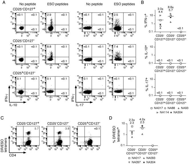
Spontaneous CD4(+) T-cell responses to the tumor-specific antigen NY-ESO-1 (ESO) are frequently found in patients with epithelial ovarian cancer (EOC). If these responses are of effector or/and Treg type, however, has remained unclear. Here, we have used functional approaches together with recently developed MHC class II/ESO tetramers to assess the frequency, phenotype and function of ESO-specific cells in circulating lymphocytes from EOC patients. We found that circulating ESO-specific CD4(+) T cells in EOC patients with spontaneous immune responses to the antigen are prevalently T(H)1 type cells secreting IFN-γ but no IL-17 or IL-10 and are not suppressive. We detected tetramer(+) cells ex vivo, at an average frequency of 1:25,000 memory cells, that is, significantly lower than in patients immunized with an ESO vaccine. ESO tetramer(+) cells were mostly effector memory cells at advanced stages of differentiation and were not detected in circulating CD25(+)FOXP3(+)Treg. Thus, spontaneous CD4(+) T-cell responses to ESO in cancer patients are prevalently of T(H)1 type and not Treg. Their relatively low frequency and advanced differentiation stage, however, may limit their efficacy, that may be boosted by immunogenic ESO vaccines.


Conflict of interest statement

Competing Interests: The authors have declared that no competing interests exist.

Figures

Figure 1
Figure 1. Phenotypic assessment of memory conventional and regulatory CD4+ T-cell subsets in circulating lymphocytes of healthy donors and EOC patients.
A. CD4+ T cells were stained with anti-FOXP3, -CD25, -CD45RA and CD127 antibodies and analyzed by flow cytometry. Expression of CD25 and CD127 defines 3 populations of memory (CD45RA) CD4+ T cells: conventional CD25CD127+ and CD25CD127 and Treg CD25+CD127 (left dot plot, numbers correspond to the proportion of each subset among memory CD4+ T cells). Histograms show the expression of FOXP3 in the defined memory CD4+ T-cell subsets. B. The proportion of conventional CD25CD127+ and CD25CD127 and Treg CD25+CD127 subsets, defined in A, among memory CD4+ T cells of healthy donors (HD, n = 27) and patients (P, n = 18).
Figure 2
Figure 2. Functional assessment of memory conventional and regulatory CD4+ T-cell subsets in circulating lymphocytes of healthy donors and EOC patients.
Ex vivo-sorted memory conventional, CD25CD127+ and CD25CD127, and Treg, CD25+CD127, populations from healthy donors (HD, n = 12) and patients (P, n = 12) were stimulated in vitro and day 12 cultures were assessed for IFN-γ, IL-10 and IL-17 production, following stimulation with PMA and ionomycin, in a 4-h intracellular cytokine secretion assay and analyzed by flow cytometry. Dot plots for one donor are shown in A and data for all healthy donors and patients are summarized in B. Statistical analyses were performed using a standard two-tailed t-test.
Figure 3
Figure 3. Assessment of ESO-specific antibody responses in sera of EOC patients.
A. The presence of ESO-specific Ab in patients' sera was assessed by ELISA. Sera were initially assessed at a 1∶100 dilution on rESO-coated or on control uncoated plates. Sera were scored as ESO Ab+ if the optical density (OD) values obtained on rESO-coated plates were both at least 3 folds higher than those obtained for the same sample on uncoated plates and higher than the mean+6xSD of OD values obtained with sera from healthy individuals on rESO-coated plates (n = 53, mean+6xSD  = 750, data not shown). B. Serial dilutions of ESO Ab+ sera were tested on rESO-coated plates. Serum titer was calculated as the serum dilution yielding 50% of maximal OD.
Figure 4
Figure 4. Assessment of ESO-specific cells in circulating memory CD4+ T-cell subsets of EOC patients.
Memory conventional, CD25CD127+ and CD25CD127, and Treg, CD25+CD127, populations were sorted ex vivo from CD4+ T cells of ESO Ab+ patients and stimulated in vitro with a pool of long overlapping peptides spanning the full length ESO sequence. A and B. Day 12 cultures were assessed for IFN-γ, IL-10 and IL-17 production in a 4-h intracellular cytokine staining assay following stimulation in the absence or presence of the ESO peptide pool. Dot plots for one patient are shown in A and data obtained for all patients are summarized in B. C and D. Day 12 cultures were stained with DR52b/ESO119–143 (NA017, NA093 and NA097) or DR4/ESO119–143 (NA304) tetramers and anti-CD4 mAb and analyzed by flow cytometry. Dot plots for one patient are shown in C and data for all patients are summarized in D.
Figure 5
Figure 5. Assessment of the suppressive activity of ESO-specific CD4+ T-cell polyclonal populations.
ESO-specific cells were isolated from peptide-stimulated CD25CD127+ and CD25CD127 CD4+ T-cell cultures (Figure 4) by tetramer-guided (NA017) or IFN-γ-guided (NA114) flow cytometry cell sorting and expanded in vitro. The resulting polyclonal cultures contained >80% ESO-specific cells. A. ESO-specific polyclonal cultures as well as polyclonal cultures from the ESO fraction and control cultures of in vitro-expanded conventional memory CD4+ T cells (M) and memory Treg (MTreg), isolated ex vivo from healthy individuals, were stained with FOXP3-specific mAb and analyzed by flow cytometry. Numbers in dot plots correspond to the mean fluorescence intensity (MFI) of FOXP3 staining. B. The suppressive activity of ESO-specific and control polyclonal populations was assessed by co-culture with CFSE-labeled conventional CD4+ T cells, at a responder:suppressor ratio of 1∶1, in the presence of irradiated monocytes and PHA. Dot plots show the CFSE-dilution profile in the absence of test population (left) and in the presence of the indicated test populations. Numbers in histograms correspond the percentage of undivided cells. Results corresponding to the calculated% suppression are shown for all tested populations.
Figure 6
Figure 6. Ex vivo assessment of ESO-specific CD4+ T cells using DR52b/ESO tetramers.
CD4+ T cells from DR52b+ healthy donors (HD) and patients were stained ex vivo with DR52b/ESO119–143 tetramers and mAb specific for CD45RA, CD25, CD127, CCR7 and CD27 and analyzed by flow cytometry. A. Dot plots for one HD and one EOC patient are shown. Numbers in dot plots correspond to the percentage of tetramer+ cells among CD45RA memory cells. Data for all EOC patients with spontaneous immune responses to ESO (S) are shown in comparison to the frequency (mean ± SD) of ESO tetramer+ cells in post-vaccine samples from patients having received a recombinant ESO vaccine (V) . B. Dot plots show the expression of CD25 and CD127 in total memory cells and in tetramer+ cells of one EOC patient. Data corresponding to the proportion of conventional, CD25CD127+ and CD25CD127, and Treg, CD25+CD127, populations within tetramer+ cells for all EOC patients (S) are summarized and compared to the proportion (mean ± SD) of these populations in vaccine-induced tetramer+ cells (V). C. Dot plots show the expression of CCR7 and CD27 in total memory cells and in tetramer+ cells of one EOC patient. Data corresponding to the proportion of CCR7+, CCR7CD27+ and CCR7CD27 populations within tetramer+ cells for all EOC patients (S) are summarized and compared to the proportion (mean ± SD) of these populations in vaccine-induced tetramer+ cells (V). Statistical analyses were performed using a standard two-tailed t-test.

References

    1. Kennedy R, Celis E. Multiple roles for CD4+ T cells in anti-tumor immune responses. Immunol Rev. 2008;222:129–144. - PubMed
    1. Muranski P, Restifo NP. Adoptive immunotherapy of cancer using CD4(+) T cells. Curr Opin Immunol. 2009;21:200–208. - PMC - PubMed
    1. Curiel TJ, Coukos G, Zou L, Alvarez X, Cheng P, et al. Specific recruitment of regulatory T cells in ovarian carcinoma fosters immune privilege and predicts reduced survival. Nat Med. 2004;10:942–949. - PubMed
    1. Sato E, Olson SH, Ahn J, Bundy B, Nishikawa H, et al. Intraepithelial CD8+ tumor-infiltrating lymphocytes and a high CD8+/regulatory T cell ratio are associated with favorable prognosis in ovarian cancer. Proc Natl Acad Sci U S A. 2005;102:18538–18543. - PMC - PubMed
    1. Hiraoka N, Onozato K, Kosuge T, Hirohashi S. Prevalence of FOXP3+ regulatory T cells increases during the progression of pancreatic ductal adenocarcinoma and its premalignant lesions. Clin Cancer Res. 2006;12:5423–5434. - PubMed

Publication types

MeSH terms