. 2011;6(7):e22931.
doi: 10.1371/journal.pone.0022931. Epub 2011 Jul 28.

Rab18 dynamics in adipocytes in relation to lipogenesis, lipolysis and obesity

Affiliations

Rab18 dynamics in adipocytes in relation to lipogenesis, lipolysis and obesity

Marina R Pulido et al. PLoS One. 2011.

Abstract

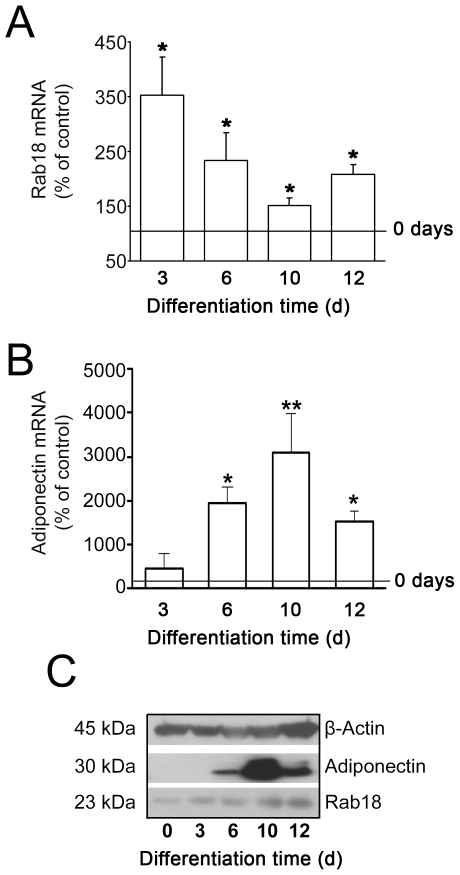
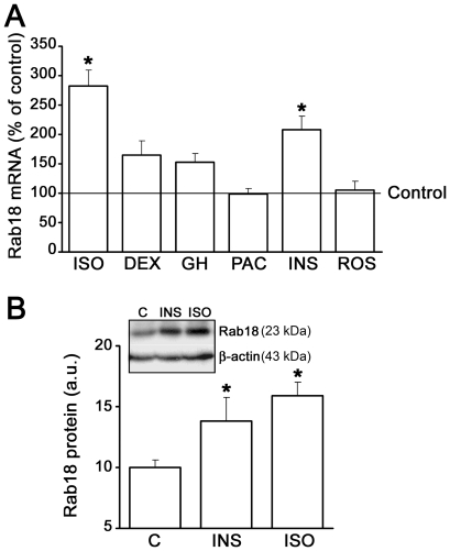
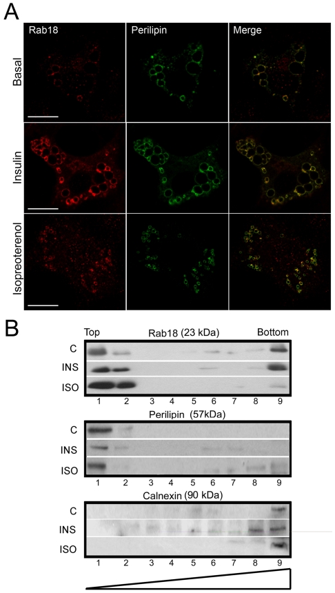
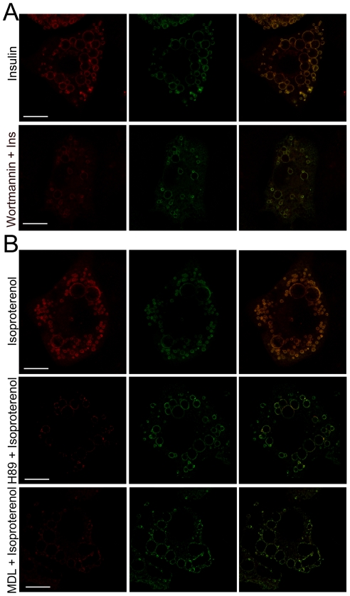
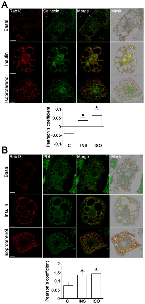
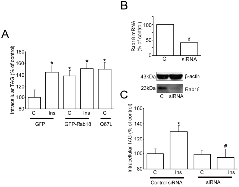
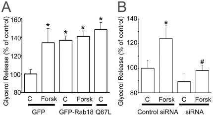
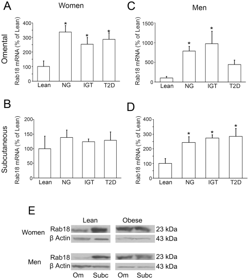
Lipid droplets (LDs) are organelles that coordinate lipid storage and mobilization, both processes being especially important in cells specialized in managing fat, the adipocytes. Proteomic analyses of LDs have consistently identified the small GTPase Rab18 as a component of the LD coat. However, the specific contribution of Rab18 to adipocyte function remains to be elucidated. Herein, we have analyzed Rab18 expression, intracellular localization and function in relation to the metabolic status of adipocytes. We show that Rab18 production increases during adipogenic differentiation of 3T3-L1 cells. In addition, our data show that insulin induces, via phosphatidylinositol 3-kinase (PI3K), the recruitment of Rab18 to the surface of LDs. Furthermore, Rab18 overexpression increased basal lipogenesis and Rab18 silencing impaired the lipogenic response to insulin, thereby suggesting that this GTPase promotes fat accumulation in adipocytes. On the other hand, studies of the β-adrenergic receptor agonist isoproterenol confirmed and extended previous evidence for the participation of Rab18 in lipolysis. Together, our data support the view that Rab18 is a common mediator of lipolysis and lipogenesis and suggests that the endoplasmic reticulum (ER) is the link that enables Rab18 action on these two processes. Finally, we describe, for the first time, the presence of Rab18 in human adipose tissue, wherein the expression of this GTPase exhibits sex- and depot-specific differences and is correlated to obesity. Taken together, these findings indicate that Rab18 is involved in insulin-mediated lipogenesis, as well as in β-adrenergic-induced lipolysis, likely facilitating interaction of LDs with ER membranes and the exchange of lipids between these compartments. A role for Rab18 in the regulation of adipocyte biology under both normal and pathological conditions is proposed.


Conflict of interest statement

Competing Interests: The authors have declared that no competing interests exist.

Figures

Figure 1
Figure 1. Rab18 expression during differentiation of 3T3-L1 cells into adipocytes.
(A) Quantitative RT-PCR analysis of Rab18 mRNA levels in 3T3-L1 cells exposed to a hormonal differentiation cocktail for 0, 3, 6, 10 and 12 days. Gene expression was expressed as the ratio of target gene concentration to the concentration of a housekeeping gene, the 18S rRNA (2.0×10⁸±7.7×10⁷ and 3.2×10¹³±2.2×10¹² Rab18 and 18S cDNA copies, respectively, per 0.1 µg total RNA in non-differentiated cells). Three independent experiments ± SEM were tested for significance using the Newman-Keul's comparison test. *, P<0.05 vs. non-differentiated cells. (B) To monitor adipogenesis, adiponectin mRNA transcripts (1.6×10⁸±1.3×10⁸ cDNA copies/0.1 µg total RNA in non-differentiated cells) were also quantified. (C) Representative immunoblots from 3 independent analyses of Rab18 and adiponectin protein content in 3T3-L1 cell extracts during differentiation. β-actin immunosignal was used as reference for protein charge.
Figure 2
Figure 2. Regulation of Rab18 gene expression and protein content in 3T3-L1 adipocytes.
(A) 3T3-L1 adipocytes were treated with 10 µM isoproterenol (ISO), 100 nM dexamethasone (DEX), 10 nM GH, 4.8 nM PACAP (PAC), 100 nM insulin (INS), or 10 µM rosiglitazone (ROS) for 24 h, and Rab18 mRNA levels were evaluated by quantitative RT-PCR. 18S rRNA expression was used as internal control (1.7×10⁹±9.2×10⁸ and 1.1×10¹²±1.5×10¹¹ Rab18 and 18S cDNA copies, respectively, per 0.1 µg total RNA in untreated cells). Data represent the average ± SEM of 3 independent experiments. *, P<0.05 vs. untreated cells. . (B) Quantification of Rab18 band intensity in extracts from 3T3-L1 adipocytes cultured in the presence or absence of 100 nM insulin (INS) or 10 µM isoproterenol (ISO) for 24 h s. β-actin immunostaining was used as loading control. Data are expressed as the mean ± SEM of 3 independent experiments. *, P<0.05 vs. untreated cells. A representative immunoblot of Rab18 and β-actin is shown.
Figure 3
Figure 3. Rab18 subcellular localization in 3T3-L1 adipocytes under insulin or isoproterenol stimulation.
(A) Confocal microscope images of 3T3-L1 cells under basal conditions (top panels) or challenged with 100 nM insulin (middle panels) or 10 µM isoproterenol (bottom panels) for 4 h and co-immunostained for Rab18 (red) and the LD-associated protein perilipin (green). Colocalization of the two immunosignals can be seen in the most right panels (yellow). Scale bars, 5 µm. (B) Immunoblots of sucrose gradients from 3T3-L1 cells under non-stimulated conditions and after 4-h treatments with 100 nM insulin and 10 µM isoproterenol. Perilipin and calnexin immunoreactivities were used as markers of LD-enriched fractions (light fractions) and microsomal fractions (heavy fractions), respectively. One representative experiment out of three with similar results is shown.
Figure 4
Figure 4. Intracellular signaling pathways mediating the effect of insulin and isoproterenol on Rab18 association with LDs.
(A) Representative confocal images of 3T3-L1 adipocytes treated with 100 nM insulin for 4 h in the absence (top panels) or presence of the PI3K blocker wortmannin (1 µM). (B) Representative confocal images of 3T3-L1 adipocytes treated with 10 µM isoproterenol for 4 h in the absence (top panels) or presence of the PKA blocker H89 (1 µM; middle panels) or the adenylate cyclase blocker MDL 12,330 (1 µM; bottom panels). After treatment, cells were co-immunostained for Rab18 (red) and perilipin (green). Colocalization of the two proteins is shown in the images on the far right (yellow). Scale bars, 5 µm.
Figure 5
Figure 5. Effects of insulin and isoproterenol on Rab18 localization in relation to the ER.
Representative confocal images of 3T3-L1 adipocytes under basal conditions or treated with 100 nM insulin or 10 µM isoproterenol for 4 h. After treatment, cells were co-immunostained for Rab18 (red) and the ER markers calnexin (A) or protein disulfure isomerase (B). In all cases, regions of interest containing a group of Rab18-positive LDs are presented. The colocalization channel was isolated using Imaris 6.4 (Bitplane) and shown alone in the images on the far right. Scale bars, 2 µm. To quantify the degree of colocalization, Pearson' coefficients were calculated for each experimental condition and represented as the mean ± SEM of, at least, 10 cells per experimental group. *, P<0.05 vs. untreated cells.
Figure 6
Figure 6. Effect of Rab18 overexpression and silencing on insulin-induced lipogenesis.
(A) The effect of Rab18 overexpression on basal and insulin-induced lipogenic activity in 3T3-L1 adipocytes was assessed by transfecting cells with GFP-tagged versions of wild-type Rab18 or the constitutively active mutant Rab18(Q67L) for 48–72 h. In parallel, a group of cells (controls) were transfected a vector expressing GFP alone. GFP fluorescence served to confirm transfection efficiencies higher than 90% for each experiment prior to further experimental manipulation. After 4-h treatment, cells were lysed and intracellular TAG content was quantified. TAG content was normalized to total protein content and expressed as the mean ± SEM of 6 independent experiments. (B) Rab18 silencing was achieved by transfecting 3T3-L1 cells with a mouse Rab18 specific siRNA. As control, a group of cells were transfected with a scramble negative control siRNA. Rab18 knock down was confirmed by quantitative RT-PCR and Western blot. (C) After 72 h, cells were challenged to 100 nM insulin for 4 h, harvested and lysed. TAG was content quantified as indicated above and expressed as the mean ± SEM of 6 independent experiments. *, P<0.05 and **, P<0.01 vs. untreated control cells. #, P<0.05 vs. insulin-treated control cells.
Figure 7
Figure 7. Effect of Rab18 overexpression and silencing on forskolin-induced lipolysis.
(A) The effect of Rab18 overexpression on basal and forskolin-induced lipolysis was evaluated by transfecting 3T3-L1 adipocytes with GFP-tagged, wild-type Rab18 or the constitutively active mutant Rab18(Q67L) for 48–72 h. In each experiment, a group of cells (controls) were transfected a vector expressing GFP alone. GFP fluorescence served to confirm transfection efficiencies higher than 90% for each experiment prior to further experimental manipulation. After 30 min in the presence or absence of forskolin, the culture medium was collected and glycerol release measured with Free Glycerol Reagent kit (Sigma-Aldrich). Glycerol concentration in the medium was normalized to total protein and expressed as the mean ± SEM of 5 independent experiments. (B) Rab18 silencing was achieved by transfecting 3T3-L1 cells with a mouse Rab18 specific siRNA. After 72 h, cells were challenged with 50 µM forskolin for 30 min. Glycerol release was quantified as indicated above and expressed as the mean ± SEM of 5 independent experiments. *, P<0.05 vs. untreated control cells. #, P<0.05 vs. forskolin-treated control cells.
Figure 8
Figure 8. Assessment of Rab18 expression levels in human adipose tissue in relation to obesity and insulin resistance.
Omental and subcutaneous adipose tissue samples were obtained from the abdominal region of women (A and B) and men (C and D) with different degrees of obesity and/or insulin-resistance: lean, obese normoglycemic (NG), obese with impaired glucose tolerance (IGT) and obese type 2 diabetic (T2D) patients. After removal, tissue samples were processed for total RNA extraction. Rab18 expression was evaluated by quantitative RT-PCR using specific primers for human Rab18. The expression of human 18S rRNA in each sample was evaluated as an internal housekeeping gene. Absolute cDNA copy number for lean patients were 2.1×10⁵±1.7×10⁵ Rab18 copies and 6.6×10¹⁰±5.5×10¹⁰ 18S copies in omental fat from women, 5.9×10⁴±3.1×10⁴ Rab18 copies and 5.7×10¹⁰±1.5×10¹⁰ 18S copies in omental fat from men, 4.9×10⁴±3.2×10⁴ Rab18 copies and 1.1×10¹⁰±6.9×10⁹ 18S copies in subcutaneous fat from women, and 5.2×10⁴±2.0×10⁴ Rab18 copies and 2.8×10¹⁰±1.3×10⁹ 18S copies in subcutaneous fat from men. *, P<0.05 vs. lean subjects. (E) Rab18 protein content in omental and subcutaneous adipose tissue samples from lean and obese women and men. In each blot, the β-actin immunosignal from the same sample was used as internal control.

Similar articles

Cited by

References

    1. Jaworski K, Sarkadi-Nagy E, Duncan RE, Ahmadian M, Sul HS. Regulation of triglyceride metabolism. IV. Hormonal regulation of lipolysis in adipose tissue. Am J Physiol Gastrointest Liver Physiol. 2007;293:G1–4. - PMC - PubMed
    1. Kersten S. Mechanisms of nutritional and hormonal regulation of lipogenesis. EMBO Rep. 2001;2:282–286. - PMC - PubMed
    1. Langin D. Adipose tissue lipolysis as a metabolic pathway to define pharmacological strategies against obesity and the metabolic syndrome. Pharmacol Res. 2006;53:482–491. - PubMed
    1. Ahmadian M, Duncan RE, Sul HS. The skinny on fat: lipolysis and fatty acid utilization in adipocytes. Trends Endocrinol Metab. 2009;20:424–428. - PMC - PubMed
    1. Ahmadian M, Wang Y, Sul HS. Lipolysis in adipocytes. Int J Biochem Cell Biol. 2010;42:555–559. - PMC - PubMed

Publication types

MeSH terms