. 2011;6(7):e22979.
doi: 10.1371/journal.pone.0022979. Epub 2011 Jul 29.

The X-linked tumor suppressor TSPX interacts and promotes degradation of the hepatitis B viral protein HBx via the proteasome pathway

Affiliations

The X-linked tumor suppressor TSPX interacts and promotes degradation of the hepatitis B viral protein HBx via the proteasome pathway

Tatsuo Kido et al. PLoS One. 2011.

Abstract

Hepatitis B virus (HBV) infection is a major risk for hepatocellular carcinoma (HCC), and it is a serious global health problem with two billion people exposed to it worldwide. HBx, an essential factor for viral replication and a putative oncoprotein encoded by the HBV genome, has been shown to promote oncogenic properties at multiple sites in HBV-infected liver cells. The expression level of HBx closely associates with the development and progression of HCC, therefore the mechanism(s) regulating the stability of HBx is important in oncogenesis of HBV-infected cells. We demonstrate that the X-linked tumor suppressor TSPX enhances the degradation of HBx through the ubiquitin-proteasome pathway. TSPX interacts with both HBx and a proteasome 19S lid subunit RPN3 via its C-terminal acidic tail. Most importantly, over-expression of RPN3 protects HBx from, and hence acts as a negative regulator for, proteasome-dependent degradation. TSPX abrogates the RPN3-depedent stabilization of HBx, suggesting that TSPX and RPN3 act competitively in regulation of HBx stability. Since mutation and/or epigenetic repression of X-located tumor suppressor gene(s) could significantly predispose males to human cancers, our data suggest that TSPX-induced HBx degradation could play key role(s) in hepatocarcinogenesis among HBV-infected HCC patients.


Conflict of interest statement

Competing Interests: The authors have declared that no competing interests exist.

Figures

Figure 1
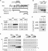
Figure 1. TPSX stimulates degradation of HBx in a proteasome dependent manner.
(A) Structure of TSPY, TSPX, and schematic representation of the truncated mutants of TSPX used in present study. (B) Effect of co-expression with TSPX on HBx stability in mammalian cells. 293T cells were co-transfected with HA-HBx expression vector (0.2 µg/well) in the presence or absence of TSPX[full] or FLAG-TSPX[ΔPro] expression vector (0.025, 0.1 µg/well). DsRed-V5 expression vector (0.1 µg/well) was co-transfected as the internal control for monitoring the transfection efficiency. Forty-eight hours after transfection, cells were lysed and analyzed by Western-blot (immuno-blot = IB) using indicated antibodies. (C) Effect of a proteasome inhibitor MG132 on TSPX-enhanced HBx degradation. 293T cells and HuH-7 cells were co-transfected with HA-HBx expression vector (0.2 µg/well) in the presence or absence of FLAG-TSPX[ΔPro] expression vector (0.025, 0.1 µg/well). DsRed-V5 expression vector (0.1 µg/well) was co-transfected similarly as above. Twenty-four hours after transfection, cells were treated with vehicle (DMSO) or 25 µM MG132 for additional 24 h. Cells were lysed and analyzed by Western-blot using indicated antibodies. (D) Interaction of TSPX and HBx in mammalian cells. HA-HBx expression vector was co-transfected into 293T cells with expression vector of FLAG-epitope tagged TSPX mutants. Twenty-four hours after transfection, cells were treated with 20 µM MG132 for additional 24 h. Coimmunoprecipitation was performed with anti-FLAG antibody, and immunoprecipitated complexes (co-IP) were analyzed by Western blot using anti-HA and anti-FLAG antibodies. One percent of each lysate (input) was analyzed in parallel as a transfection control. (E) Mapping of the functional domain for stimulation of HBx-degradation. HA-HBx expression vector (0.1 µg/well) was co-transfected into 293T cells with FLAG-TSPX[ΔPro] (0.05, 0.1, 0.2 µg/well), FLAG-TSPX[ΔProΔC] (0.05, 0.1, 0.2 µg/well), FLAG-TSPX[Tail-L] (0.05, 0.1, 0.2 µg/well) or FLAG-TSPX[Tail-S] (0.05, 0.1, 0.2 µg/well), and analyzed as described above. The results indicate that the D/E-rich C-terminal region is sufficient to promote HBx degradation.
Figure 2
Figure 2. Endogenously expressed TSPX enhances HBx degradation in 293T cells.
(A) 293T cells express endogenous TSPX. Total RNA isolated from 293T cells were analyzed by RT-PCR with primer pairs for TSPX[538–693] and GAPDH using a standard technique. Control, PCR product with p3×FLAG-TSPX[ΔPro]; +RT, with reverse transcriptase; -RT, without reverse transcriptase. (B) Repression of endogenous TSPX increased the expression of HA-HBx in 293T cells. HA-HBx expression vector (50 ng/well) and DsRed-V5 expression vector (50 ng/well) were co-transfected into 293T cells with either control siRNA (4 pmol/well) or TSPX siRNA (4 pmol/well). Forty-eight hours after transfection, cells were lysed and analyzed by Western-blot using anti-V5 and anti-HA antibodies. Treatment with TSPX siRNA resulted in significant increase in HA-HBx expression (2.8 fold). (C) Effect of TSPX siRNA on TSPX expression. FLAG-TSPX[ΔPro] expression vector (0.1 µg/well) was co-transfected into 293T cells with control siRNA (4 pmol/well) or TSPX siRNA (4 pmol/well). Forty-eight hours after transfection, cells were lysed and analyzed by western-blot using anti-FLAG antibody. TSPX siRNA significantly decreased the protein level of FLAG-TSPX[ΔPro].
Figure 3
Figure 3. TSPX mediates HBx degradation through ubiquitin-proteasome pathway.
Ubiquitin-activating enzyme inhibitor PYR-41 inhibits TSPX-mediated enhancement of HBx-degradation. HA-HBx expression vector (0.2 µg/well) and DsRed-V5 expression vector (0.1 µg/well) were co-transfected into 293T cells with FLAG-TSPX[ΔPro] (0.05 µg/well). Twenty-four hours after transfection, cells were treated with either vehicle (DMSO) or 50 µM PYR-41 for additional 24 h. Cell lysates were analyzed by Western blot using anti-HA, anti-V5 and anti-FLAG antibodies. Treatment with PYR-41 significantly increased the HA-HBx even in the presence of TSPX.
Figure 4
Figure 4. TSPX interacts with RPN3, and abrogates the protective effect of RPN3 on HBx-degradation.
(A) Structure of RPN3 and schematic representation of the truncated mutant of RPN3. The position of PCI/PINT domain is as marked. N-terminal truncated RPN3 (residues 128–534 a.a., termed as RPN3[ΔN]) was cloned into pCMV-Myc vector to express a Myc epitope-tagged product. (B) Interaction of TSPX and RPN3 in mammalian cells. Myc-RPN3[ΔN] was co-transfected with p3×FLAG-CMV7 (FLAG control), FLAG-TSPX[ΔPro], FLAG-TSPX[Tail-L] or FLAG-TSPX[ΔProΔC] into 293T cells. Immunoprecipitations were carried out as before using anti-FLAG antibody. Immunoblots were probed with anti-Myc and anti-FLAG antibodies. The immunoblots indicate that human RPN3 co-immunoprecipitates with human TSPX. (C) RPN3 did not interfere the interaction between TSPX and HBx. HA-HBx and FLAG-TSPX[ΔPro] expression vectors were co-transfected with or without Myc-RPN3[ΔN] vector into 293T cells. Immunoprecipitations were carried out using anti-FLAG antibody. Immunoblots were probed with anti-HA, anti-Myc and anti-FLAG antibodies, respectively. The results indicate that interaction between HA-HBx and FLAG-TSPX was not affected by co-expression of Myc-RPN3[ΔN]. (D) Over-expression of RPN3 protected HBx from protein-degradation, and TSPX overcomes the protective effect of RPN3. 293T cells were co-transfected with HA-HBx (0.2 µg/well), Myc-RPN[ΔN] (0.05, 0.1 µg/well), RPN3[full] (0.05, 0.1 µg/well) and/or FLAG-TSPX[ΔPro] (0.05 µg/well) as indicated in the figure. pcDNA-DsRed-V5 expression vector (0.1 µg/well) was co-transfected as the internal control for monitoring the transfection efficiency. Cells were lysed 48 h after transfection, and analyzed by Western blot using indicated antibodies. Although co-transfection of RPN3[full] or RPN3[ΔN] resulted in the increase of HA-HBx, TSPX significantly decreased the level of HBx even in the presence of RPN3. (E) Over-expression of Myc-RPN3[ΔN] did not affect on the transcription of HA-HBx. 293T cells were co-transfected with HA-HBx (0.2 µg/well), Myc-RPN[ΔN] (0.1 µg/well), and/or FLAG-TSPX[ΔPro] (0.05 µg/well) as indicated in figure. pcDNA-DsRed-V5 (0.1 µg/well) was co-transfected as the internal control for monitoring the transfection efficiency. Twenty-four hours after transfection, cells were treated with either vehicle (DMSO) or 25 µM MG132 for additional 24 h. Cells were lysed and analyzed by Western blot using anti-HA, anti-V5, anti-Myc, and anti-FLAG antibodies. Co-transfection of Myc-RPN3[ΔN] increased HA-HBx (DMSO). No significant difference was observed in HA-HBx level in the cells treated with MG132 (+MG132).
Figure 5
Figure 5. A model illustrating the potential roles of TSPX and RPN3 as regulators of ubiquitin-proteasome dependent HBx degradation.
TSPX enhances the HBx degradation by recruiting HBx to proteasome complex, and inhibiting the protective function of PRN3 on HBx-degradation. E1-E3 indicates the ubiquitylation cascade including E1, E2, and E3 enzymes. MG132-responsive and PYR-41-responsive sites are also indicated.

Similar articles

Cited by

References

    1. Park YJ, Luger K. Structure and function of nucleosome assembly proteins. Biochem Cell Biol. 2006;84:549–558. - PubMed
    1. Muto S, Senda M, Akai Y, Sato L, Suzuki T, et al. Relationship between the structure of SET/TAF-Ibeta/INHAT and its histone chaperone activity. Proc Natl Acad Sci U S A. 2007;104:4285–4290. - PMC - PubMed
    1. Ozbun LL, Martinez A, Jakowlew SB. Differentially expressed nucleolar TGF-beta1 target (DENTT) shows tissue-specific nuclear and cytoplasmic localization and increases TGF-beta1-responsive transcription in primates. Biochim Biophys Acta. 2005;1728:163–180. - PubMed
    1. Scully RE. Gonadoblastoma. A review of 74 cases. Cancer. 1970;25:1340–1356. - PubMed
    1. Page DC. Hypothesis: a Y-chromosomal gene causes gonadoblastoma in dysgenetic gonads. Development. 1987;101(Suppl):151–155. - PubMed

Publication types

MeSH terms