

Trauma hemorrhagic shock-induced lung injury involves a gut-lymph-induced TLR4 pathway in mice
- PMID: 21829592
- PMCID: PMC3150139
- DOI: 10.1371/journal.pone.0014829
Trauma hemorrhagic shock-induced lung injury involves a gut-lymph-induced TLR4 pathway in mice
Abstract
Background: Injurious non-microbial factors released from the stressed gut during shocked states contribute to the development of acute lung injury (ALI) and multiple organ dysfunction syndrome (MODS). Since Toll-like receptors (TLR) act as sensors of tissue injury as well as microbial invasion and TLR4 signaling occurs in both sepsis and noninfectious models of ischemia/reperfusion (I/R) injury, we hypothesized that factors in the intestinal mesenteric lymph after trauma hemorrhagic shock (T/HS) mediate gut-induced lung injury via TLR4 activation.
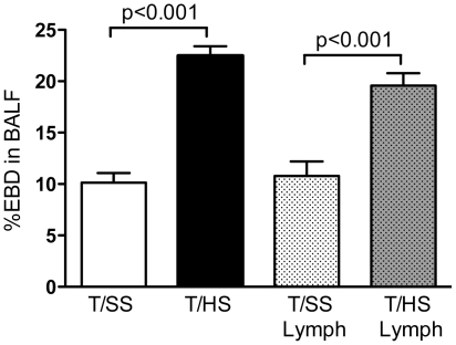
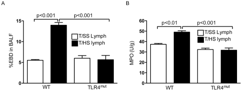
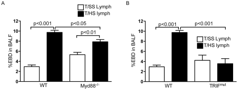
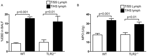
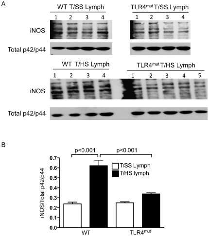
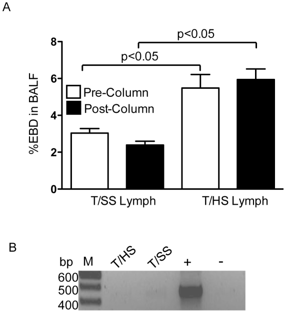
Methods/principal findings: The concept that factors in T/HS lymph exiting the gut recreates ALI is evidenced by our findings that the infusion of porcine lymph, collected from animals subjected to global T/HS injury, into naïve wildtype (WT) mice induced lung injury. Using C3H/HeJ mice that harbor a TLR4 mutation, we found that TLR4 activation was necessary for the development of T/HS porcine lymph-induced lung injury as determined by Evan's blue dye (EBD) lung permeability and myeloperoxidase (MPO) levels as well as the induction of the injurious pulmonary iNOS response. TRIF and Myd88 deficiency fully and partially attenuated T/HS lymph-induced increases in lung permeability respectively. Additional studies in TLR2 deficient mice showed that TLR2 activation was not involved in the pathology of T/HS lymph-induced lung injury. Lastly, the lymph samples were devoid of bacteria, endotoxin and bacterial DNA and passage of lymph through an endotoxin removal column did not abrogate the ability of T/HS lymph to cause lung injury in naïve mice.
Conclusions/significance: Our findings suggest that non-microbial factors in the intestinal mesenteric lymph after T/HS are capable of recreating T/HS-induced lung injury via TLR4 activation.
Conflict of interest statement
Figures
References
-
- Deitch E, Forsythe R, Anjaria D, Livingston D, Lu Q, et al. The role of lymph factors in lung injury, bone marrow suppression, and endothelial cell dysfunction in a primate model of trauma-hemorrhagic shock. Shock. 2004;22:221–228. - PubMed
-
- Senthil M, Watkins A, Barlos D, Xu DZ, Lu Q, et al. Intravenous injection of trauma-hemorrhagic shock mesenteric lymph causes lung injury that is dependent upon activation of the inducible nitric oxide synthase pathway. Ann Surg. 2007;246:822–830. - PubMed
-
- Deitch E, Xu D, Lu Q. Gut lymph hypothesis of early shock and trauma-induced multiple organ dysfunction syndrome. J Org Dysf. 2006;2:70–79.
Publication types
MeSH terms
Substances
Grants and funding
LinkOut - more resources
Full Text Sources
Medical
Molecular Biology Databases
Research Materials
Miscellaneous
