Study on phylogenetic relationships, variability, and correlated mutations in M2 proteins of influenza virus A
- PMID: 21829678
- PMCID: PMC3149066
- DOI: 10.1371/journal.pone.0022970
Study on phylogenetic relationships, variability, and correlated mutations in M2 proteins of influenza virus A
Abstract
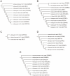
M2 channel, an influenza virus transmembrane protein, serves as an important target for antiviral drug design. There are still discordances concerning the role of some residues involved in proton transfer as well as the mechanism of inhibition by commercial drugs. The viral M2 proteins show high conservativity; about 3/4 of the positions are occupied by one residue in over 95%. Nine M2 proteins from the H3N2 strain and possibly two proteins from H2N2 strains make a phylogenic cluster closely related to 2RLF. The variability range is limited to 4 residues/position with one exception. The 2RLF protein stands out by the presence of 2 serines at the positions 19 and 50, which are in most other M2 proteins occupied by cysteines. The study of correlated mutations shows that there are several positions with significant mutational correlation that have not been described so far as functionally important. That there are 5 more residues potentially involved in the M2 mechanism of action. The original software used in this work (Consensus Constructor, SSSSg, Corm, Talana) is freely accessible as stand-alone offline applications upon request to the authors. The other software used in this work is freely available online for noncommercial purposes at public services on bioinformatics such as ExPASy or NCBI. The study on mutational variability, evolutionary relationship, and correlated mutation presented in this paper is a potential way to explain more completely the role of significant factors in proton channel action and to clarify the inhibition mechanism by specific drugs.
Conflict of interest statement
Figures


References
-
- Lamb RA, Holsinger LJ, Pinto LH. Wimmer E, editor. Receptor-Mediated Virus Entry into Cells. 1994. pp. 303–321. Cold Spring Harbor Laboratory Press, Cold Spring Harbor, 1994.
-
- Helenius A. Unpacking the incoming influenza-virus. Cell. 1992;69:577–578. - PubMed
-
- Ciampor F, Bayley PM, Nermut MV, Hirst EM, Sugrue RJ, et al. Evidence that the Amantadine-induced, M2-mediated conversion of influenza A virus hemagglutinin to the low pH conformation occurs in an acidic trans Golgi compartment. Virology. 1992;188:14–24. - PubMed
Publication types
MeSH terms
Substances
Associated data
- Actions
LinkOut - more resources
Full Text Sources
Other Literature Sources

