. 2011;6(8):e23110.
doi: 10.1371/journal.pone.0023110. Epub 2011 Aug 1.

Aurora-A interacts with AP-2α and down regulates its transcription activity

Affiliations

Aurora-A interacts with AP-2α and down regulates its transcription activity

Lihui Zou et al. PLoS One. 2011.

Abstract

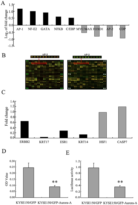
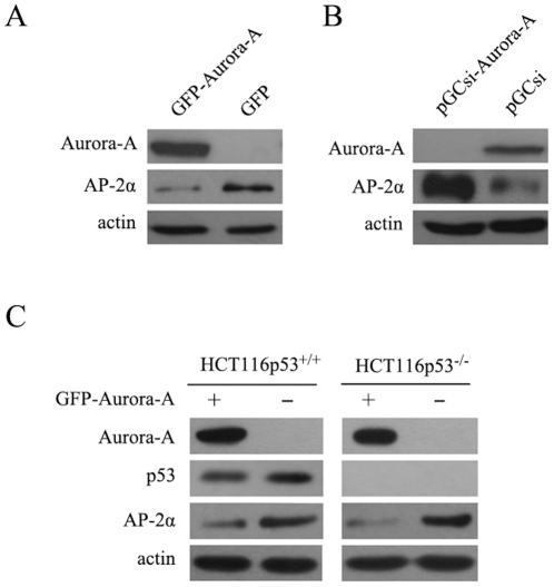
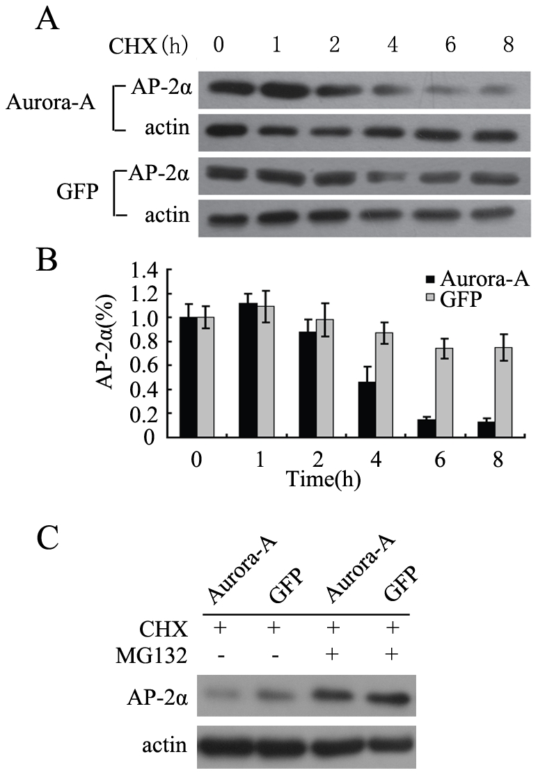
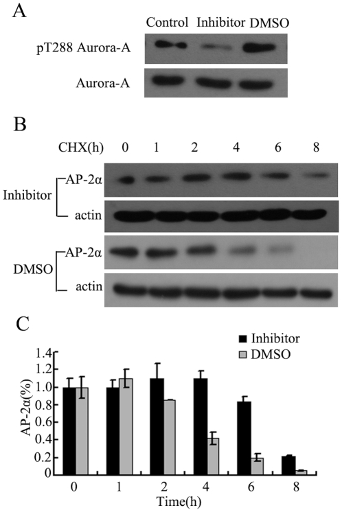
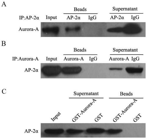
Aurora-A is a serine/threonine protein kinase and plays an important role in the control of mitotic progression. Dysregulated expression of Aurora-A impairs centrosome separation and maturation, which lead to disrupted cell cycle progression and tumorigenesis. However, the molecular mechanism by which Aurora-A causes cell malignant transformation remains to be further defined. In this report, using transcription factors array and mRNA expression profiling array, we found that overexpression of Aurora-A suppressed transcription activity of AP-2α, a tumor suppressor that is often downregulated in variety of tumors, and inhibited expression of AP-2α-regulated downstream genes. These array-based observations were further confirmed by microwell colorimetric TF assay and luciferase reporter assay. Downregulated transcription activity of AP-2α by Aurora-A was found to be associated with reduced AP-2α protein stability, which appeared to be mediated by Aurora-A enhanced ubiquitin-dependent proteasomal degradation of AP-2α protein. Interestingly, Aurora-A-mediated AP-2α degradation was likely dependent Aurora-A kinase activity since inhibition of Aurora-A kinase activity was able to rescue Aurora-A-induced degradation of AP-2α. Moreover, we defined a physical interaction between Aurora-A and AP-2α, and such interaction might bridge the suppressive effect of Aurora-A on AP-2α protein stability. These findings provide new insights into molecular mechanism by which Aurora-A acts as an oncogenic molecule in tumor occurrence and malignant development.


Conflict of interest statement

Competing Interests: The authors have read the journal's policy and have the following competing interest. Yimin Sun is an employee of CapitalBio Corporation. There are no patents, products in development or marketed products to declare. This does not alter the authors' adherence to all the PLoS ONE policies on sharing data and material, as detailed online in the guide for authors.

Figures

Figure 1
Figure 1. Transcription activity of AP-2α is downregulated by Aurora-A.
(A) The summary of the transcription factors with significant changes in their activities by transcription factors array when Aurora-A was overexpressed. (B) Fluorescence images of the transcription factors arrays hybridized with nuclear extracts from KYSE150/GFP-Aurora-A and KYSE150/GFP cells. The analysis of arrays was replicated twice, exchanging the Cy3- and Cy5- label. Magnified panels show the spots representing the activity of AP-2 transcription factor that was down-regulated upon Aurora-A overexpression. (C) The summary of the AP-2α-regulated genes with significant down-expression when Aurora-A was overexpressed by mRNA expression profiling array. The black bars represent the AP-2α-regulated genes, and the gray bars represent the genes which were not the AP-2α-regulated genes. (D) The nucleoprotein extraction of KYSE150/GFP-Aurora-A and KYSE150/GFP cells were incubated with AP-2α binding DNA double-stranded oligonucleotidic probe on multi-well plates, then anti-AP-2α antibodies and the peroxidase-conjugated anti-mouse IgG were subsequently added. At last, the samples could be read for the colorimetric detection. (E) KYSE150/GFP-Aurora-A and KYSE150/GFP cells were cotransfected with (AP-2)3-TK-LUC expression vector and a pRL-SV40 expression vector. Transcription activities were expressed as luciferase values after normalization. All experiments were performed three times and described as mean ± SD.
Figure 2
Figure 2. Regulation of AP-2α gene expression by Aurora-A.
(A) AP-2α protein levels in KYSE150/GFP-Aurora-A and KYSE150/GFP cells were analyzed using immunoblotting assay. The actin levels were measured as loading control. (B) AP-2α protein levels in EC9706/pGCsi???EC9706/pGCsi-Aurora-A cells were examined by Western Blotting assay. (C) The HCT116 p53+/+ and HCT116 p53-/- cells were transiently transfected with pEGFP-Aurora-A and control plasmids. Cells were lysed after 48h of incubation and evaluated for the expression of AP-2α by Western Blotting assay.
Figure 3
Figure 3. Overexpression of Aurora-A enhances the degradation of AP-2α.
(A) KYSE150/GFP-Aurora-A and KYSE150/GFP cells were treated with CHX respectively, and then harvested at the indicated time points. AP-2α protein level at each time point was determined by Western Blot. (B) The amounts of AP-2α were calculated by densitometry and normalized to corresponding actin levels. The column diagram represents the amount of normalized AP-2α at each time point comparing with the original levels (0 h). (C) Aurora-A enhances the proteasome-dependent degradation of AP-2α. KYSE150/GFP-Aurora-A and KYSE150/GFP cells were treated with CHX alone or combination with MG-132. AP-2α protein level was determined by Western Blot.
Figure 4
Figure 4. Aurora-A-mediated AP-2α degradation depends on Aurora-A kinase activity.
(A) KYSE150/GFP-Aurora-A cells were exposed to 1 µM of Aurora-A kinase inhibitor and cellular proteins were collected 2 hours later. Western Blot was performed with antibodies to pThr288 on Aurora-A or total Aurora-A. DMSO was used as a negative control. (B) KYSE150/GFP-Aurora-A cells were exposed to Aurora-A kinase inhibitor or DMSO for 2 hours prior to treatment with CHX. Cells were harvested at the indicated time points and analyzed by Western Blot. (C) The amounts of AP-2α were calculated by densitometry and normalized to corresponding actin levels. The column diagram represents the amount of normalized AP-2α at each time point comparing with the original levels (0 h).
Figure 5
Figure 5. Physical association of Aurora-A with AP-2α.
(A‵B) Protein extracts from KYSE150/GFP-Aurora-A cells were prepared and immunoprecipitated with anti-Aurora-A, anti-AP-2α, mouse or rabbit IgG. The immunocomplexes were analyzed by SDS-PAGE and immunoblotted with antibodies against Aurora-A and AP-2α respectively. The visualized bands were shown for Aurora-A and AP-2α. (C) Aurora-A-GST fusion protein was bound to glutathione-sepharose beads, and incubated with protein extracts. The pull-down complexes were analyzed by SDS-PAGE and immunoblotted with antibodies against AP-2α. The visualized band of 48 kDa was AP-2α.

Similar articles

Cited by

References

    1. Giet R, Prigent C. Aurora/Ipl1p-related kinases, a new oncogenic family of mitotic serine-threonine kinases. J Cell Sci. 1999;112(Pt21):3591–3601. - PubMed
    1. Bischoff JR, Plowman GD. The Aurora/Ipl1p kinase family: regulators of chromosome segregation and cytokinesis. Trends Cell Biol. 1999;9:454–459. - PubMed
    1. Fu J, Bian M, Jiang Q, Zhang C. Roles of Aurora kinases in mitosis and tumorigenesis. Mol Cancer Res. 2007;5:1–10. - PubMed
    1. Bischoff JR, Anderson L, Zhu Y, Mossie K, Ng L, et al. A homologue of Drosophila aurora kinase is oncogenic and amplified in human colorectal cancers. EMBO J. 1998;17:3052–3065. - PMC - PubMed
    1. Sen S, Zhou H, White RA. A putative serine/threonine kinase encoding gene BTAK on chromosome 20q13 is amplified and overexpressed in human breast cancer cell lines. Oncogene. 1997;14:2195–2200. - PubMed

Publication types

MeSH terms

Substances