Comparative Study
. 2011 Sep;20 Suppl 5(Suppl 5):619-25.
doi: 10.1007/s00586-011-1929-9. Epub 2011 Aug 10.

Sagittal balance analysis after pedicle subtraction osteotomy in ankylosing spondylitis

Affiliations
Comparative Study

Sagittal balance analysis after pedicle subtraction osteotomy in ankylosing spondylitis

Romain Debarge et al. Eur Spine J. 2011 Sep.

Abstract

Introduction: This is a radiographic study of ankylosing spondylitis patients with severe fixed kyphotic deformity who underwent pedicle subtraction osteotomy. Our goal was to measure and validate new angle to assess global kyphosis and to evaluate the sagittal balance after surgery. This is the first report which describes new angle to assess global kyphosis (T1-S1).

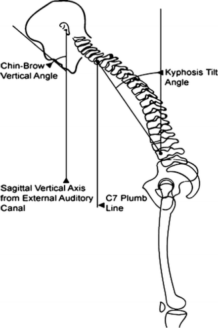
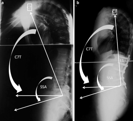
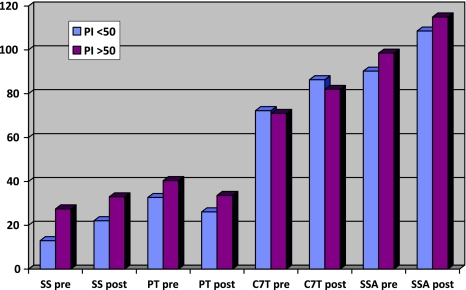
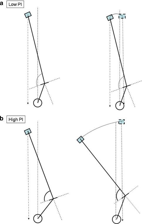
Materials and methods: Pre and postoperative controls were compared according to the Pelvic Incidence. The sagittal parameters ankylosing spondylitis patients were compared with 154 asymptomatic patients. In addition to the pelvic parameters and the C7 tilt, we used the spino-sacral angle.

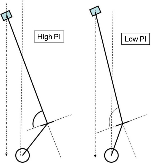
Results: Pelvic incidence in ankylosing spondylitis patients was higher than asymptomatic population (61° vs. 51°). For a same tilt of C7 for both groups, the low pelvic incidence group had a lower sacral slope and pelvic tilt and a higher global kyphosis (spino-sacral angle = 90°) than the high pelvic incidence group (spino-sacral angle = 98°). In the adult volunteers, the C7 tilt and spino-sacral angle measured, respectively, 95° and 135°. The preoperative C7 tilt measured 73° and increased to 83° (p = 0.0025). The preoperative spino-sacral angle measured 96° and increased to 113.3° (p = 0.003).

Conclusion: A low pelvic incidence pelvis has a lower sacral slope than in high pelvic incidence and can support a bigger kyphosis. All the parameters were improved by the pedicle subtraction osteotomy, but the average spinosacral angle remained lower than the control group. When C7 tilt was useful to assess the improvement of the sagittal balance, SSA allowed a better evaluation of the correction of kyphosis itself.


Figures

Fig. 1
Fig. 1
The measurement of the radiographic severity of the kyphotic deformity includes a description of the chin-brow to vertical angle; the position of the sagittal vertical axis from the external auditory canal; the C7 plumb line; and the kyphosis tilt angle
Fig. 2
Fig. 2
a The spino-sacral angle (SSA) is defined as the angle between a line from the center of C7 to the center of the sacral endplate and the sacral endplate itself. b Postoperative radiographic of the same patient. Transpedicular osteotomy of L4
Fig. 3
Fig. 3
Radiological results of the AS patients in function of the PI value. SS pre sacral slope preoperative, SS post sacral slope postoperative, PI pelvic incidence, SSA pre spino-sacral angle preoperative, SSA post spino-sacral angle postoperative, C7T pre C7 tilt preoperative, C7T post C7 tilt postoperative
Fig. 4
Fig. 4
For the same preoperative C7 plumb line, the patients with a high PI have a lower SSA (left) than those with a low PI (right)
Fig. 5
Fig. 5
a, b The correction of a same unbalanced C7 plumb line needs a smaller osteotomy in case of low grade incidence than with a high Pelvic Incidence. The strategy of correction of severe kyphosis has to take in account the level of pelvic incidence to appreciate the quantity of the osteotomy

References

    1. McMaster MJ, Coventry MB. Spinal osteotomy in ankylosing spondylitis. Technique, complications, and long-term results. Mayo Clin Proc. 1973;48:476–486. - PubMed
    1. White AA, 3rd, Panjabi MM, Thomas CL. The clinical biomechanics of kyphotic deformities. Clin Orthop Relat Res. 1977;128:8–17. - PubMed
    1. Berven SH, Deviren V, Smith JA, et al. Management of fixed sagittal plane deformity: results of the transpedicular wedge resection osteotomy. Spine. 2001;26:2036–2043. doi: 10.1097/00007632-200109150-00020. - DOI - PubMed
    1. Kim KT, Suk KS, Cho YJ, et al. Clinical outcome results of pedicle subtraction osteotomy in ankylosing spondylitis with kyphotic deformity. Spine. 2002;27:612–618. doi: 10.1097/00007632-200203150-00010. - DOI - PubMed
    1. Royen BJ, Gast A, Smit TH. Deformity planning for sagittal plane corrective osteotomies of the spine in ankylosing spondylitis. Eur Spine J. 2000;9:492–498. doi: 10.1007/s005860000183. - DOI - PMC - PubMed