

Retinal input influences the size and corticocortical connectivity of visual cortex during postnatal development in the ferret
- PMID: 21830218
- PMCID: PMC3670942
- DOI: 10.1002/cne.22738
Retinal input influences the size and corticocortical connectivity of visual cortex during postnatal development in the ferret
Abstract
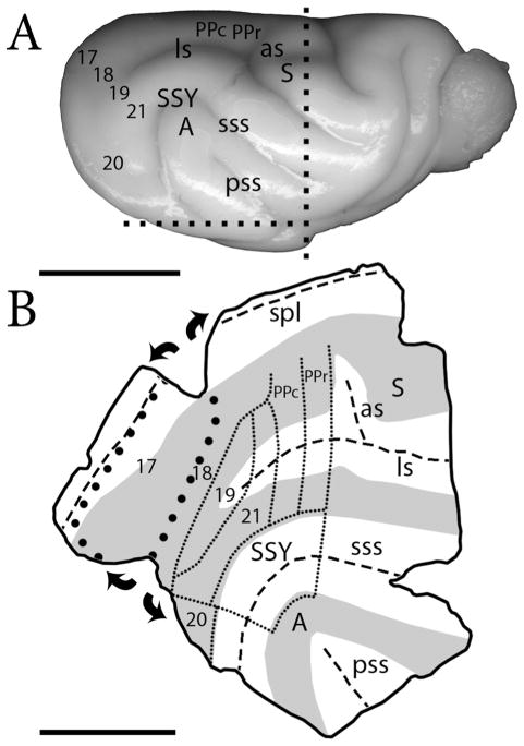
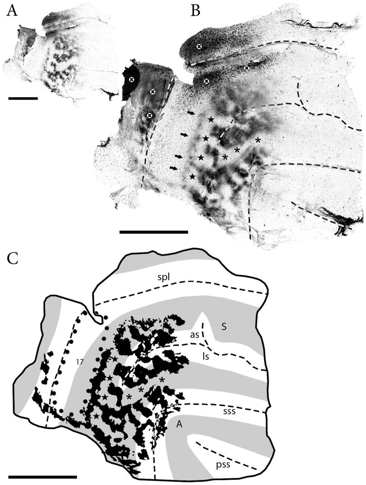
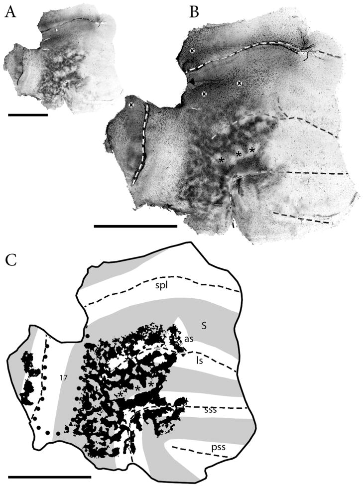
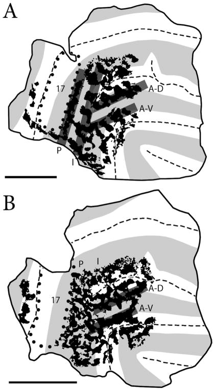
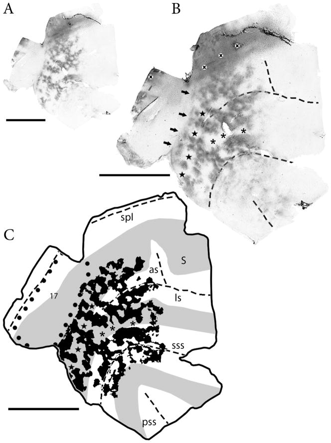
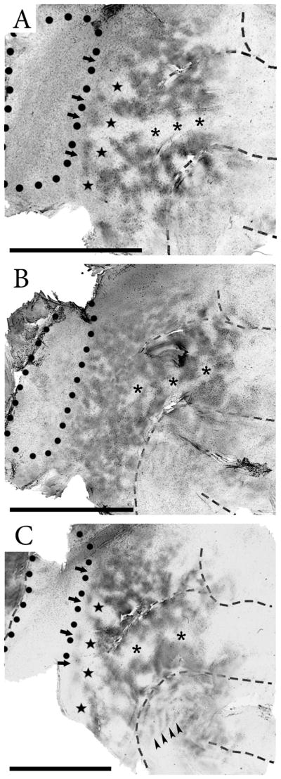
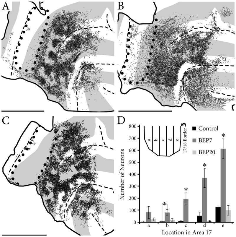
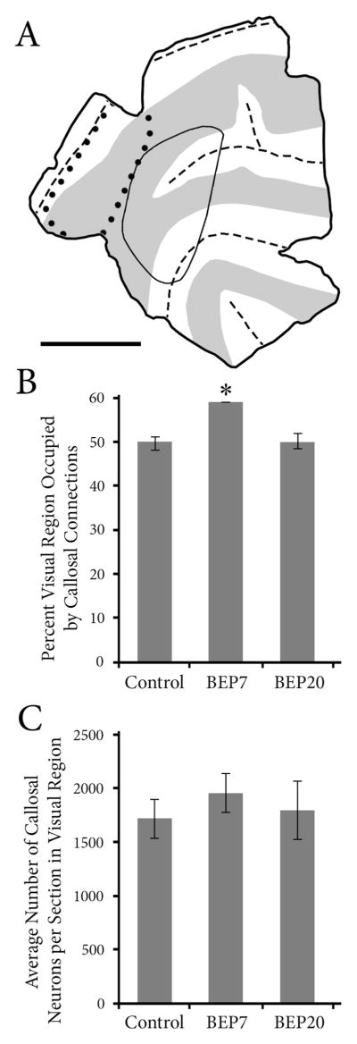
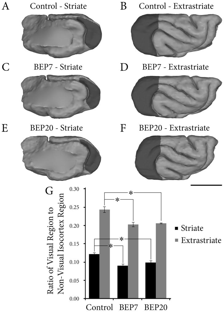
Retinal input plays an important role in the specification of topographically organized circuits and neuronal response properties, but the mechanism and timing of this effect is not known in most species. A system that shows dramatic dependence on retinal influences is the interhemispheric connection through the corpus callosum. Using ferrets, we analyzed the extent to which development of the visual callosal pattern depends on retinal influences, and explored the period during which these influences are required for normal pattern formation. We studied the mature callosal patterns in normal ferrets and in ferrets bilaterally enucleated (BE) at postnatal day 7 (P7) or P20. Callosal patterns were revealed in tangential sections from unfolded and flattened brains following multiple injections of horseradish peroxidase in the opposite hemisphere. We also estimated the effect of enucleation on the surface areas of striate and extrastriate visual cortex by using magnetic resonance imaging (MRI) data from intact brains. In BEP7 ferrets we found that the pattern of callosal connections was highly anomalous and the sizes of both striate and extrastriate visual cortex were significantly reduced. In contrast, enucleation at P20 had no significant effect on the callosal pattern, but it still caused a reduction in the size of striate and extrastriate visual cortex. Finally, retinal deafferentation had no significant effect on the number of visual callosal neurons. These results indicate that the critical period during which the eyes influence the development of callosal patterns, but not the size of visual cortex, ends by P20 in the ferret.
Copyright © 2011 Wiley Periodicals, Inc.
Figures
References
-
- Abel PL, O’Brien BJ, Olavarria JF. Organization of callosal linkages in visual area V2 of macaque monkey. J Comp Neurol. 2000;428:278–293. - PubMed
-
- Aggoun-Aouaoui D, Kiper DC, Innocenti GM. Growth of callosal terminal arbors in primary visual areas of the cat. Eur J Neurosci. 1996;8:1132–1148. - PubMed
-
- Bock AS, Kroenke CD, Taber EN, Olavarria JF. Critical period for the effects of neonatal enucleation on callosal connectivity and water diffusion anisotropy in the ferret visual system. Program No. 580.16; Society for Neuroscience Annual Meeting; 2010; San Diego, CA. 2010a.
-
- Bourdet C, Olavarria JF, Van Sluyters RC. Distribution of visual callosal neurons in normal and strabismic cats. J Comp Neurol. 1996;366:259–269. - PubMed
Publication types
MeSH terms
Grants and funding
LinkOut - more resources
Full Text Sources
