. 2011 Dec;59(12):1813-21.
doi: 10.1002/glia.21225. Epub 2011 Aug 9.

CXCR4 signaling regulates remyelination by endogenous oligodendrocyte progenitor cells in a viral model of demyelination

Affiliations

CXCR4 signaling regulates remyelination by endogenous oligodendrocyte progenitor cells in a viral model of demyelination

Kevin S Carbajal et al. Glia. 2011 Dec.

Abstract

Following intracranial infection with the neurotropic JHM strain of mouse hepatitis virus (JHMV), susceptible mice will develop widespread myelin destruction that results in pathological and clinical outcomes similar to those seen in humans with the demyelinating disease Multiple Sclerosis (MS). Partial remyelination and clinical recovery occurs during the chronic phase following control of viral replication yet the signaling mechanisms regulating these events remain enigmatic. Here we report the kinetics of proliferation and maturation of oligodendrocyte progenitor cells (OPCs) within the spinal cord following JHMV-induced demyelination and that CXCR4 signaling contributes to the maturation state of OPCs. Following treatment with AMD3100, a specific inhibitor of CXCR4, mice recovering from widespread demyelination exhibit a significant (P < 0.01) increase in the number of OPCs and fewer (P < 0.05) mature oligodendrocytes compared with HBSS-treated animals. These results suggest that CXCR4 signaling is required for OPCs to mature and contribute to remyelination in response to JHMV-induced demyelination. To assess if this effect is reversible and has potential therapeutic benefit, we pulsed mice with AMD3100 and then allowed them to recover. This treatment strategy resulted in increased numbers of mature oligodendrocytes, enhanced remyelination, and improved clinical outcome. These findings highlight the possibility to manipulate OPCs in order to increase the pool of remyelination-competent cells that can participate in recovery.


Figures

Fig. 1
Fig. 1
Immune mediated demyelination induced by intracranial infection with JHMV. (A) Following intracranial (i.c.) infection of C57BL/6 mice with JHMV, viral titers within the brain will peak between 3 and 5 days p.i. and subsequently viral replication will be reduced below levels of detection (~100 pfu g−1 tissue) (n ≥ 5 mice per time point; data is presented as average+ SD). (B) Representative coronal spinal cord section stained with luxol fast blue (LFB) and H&E depicts the characteristic ventral and lateral white matter demyelination in animals infected with JHMV at Day 35 p.i. (C) A representative light microscopy image of a toluidine blue-stained coronal section showing remyelinated (*) and demyelinated (#) axons at 35 days p.i., (scale bar= 5 μm).
Fig. 2
Fig. 2
OPCs found throughout the spinal cord proliferate and replenish mature oligodendrocyte numbers following JHMV infection. Representative immunofluorescence staining showing GSTπ-positive cells (A) in the spinal cord 35 days p.i. and a PDGFRα-positive cell (B) expressing the proliferative marker Ki67 on Day 6 p.i. Spinal cords from JHMV-infected mice were removed at defined times p.i. and (C) GST-π, (D) PDGFRα/Ki67, and (E) PDGFRα-positive cell numbers in spinal cord white matter quantified. (F) PDGFRα-positive cells were enumerated within total white matter tracts (total WM—both demyelinated and normal), normal appearing white matter (LFB-positive), and demyelinated (LFB-negative) tracts at Day 14 p.i. as described in Materials and Methods. For Panels A and B, scale bars = 20 μm. Data in C, D, E, and F represent a minimum of four mice/time point; data is presented as average ± SD; **P < 0.01.
Fig. 3
Fig. 3
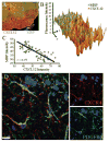
CXCL12 is expressed in areas of demyelination and OPCs express CXCR4. (A) Representative immunofluorescent image of spinal cord white matter from a mouse 35 days p.i. stained for MBP (green) and CXCL12 (red). (B) Immunofluorescence intensity plot indicating the relationship between MBP staining (green) and CXCL12 (red). (C) Regression analysis reveals increased staining for CXCL12 is associated with diminished MBP staining. Data were collected from over 50 images from three mice (Day 35 p.i.). (D) Representative confocal image of sections stained for PDGFRα and CXCR4-pS339 reveals OPCs in white matter of JHMV-infected mice at Day 35 p.i. stain positive for the active form of this receptor (>95% in all sections stained). Scale bar = 10 μm.
Fig. 4
Fig. 4
Treatment with AMD3100, a CXCR4 small molecule antagonist, in JHMV-infected mice does not affect T cell infiltration into the CNS. JHMV-infected mice were treated with either HBSS or AMD3100 for 2 weeks beginning on Day 14 p.i. and the effects on neuroinflammation evaluated after 2 weeks of treatment. (A) Representative flow dot plots of cells stained for CD4 and CD8 antigens; numbers represent frequency of positive cells within the gated population. (B) Quantitative analysis of CD4+ and CD8+ T cell infiltration into the CNS following treatment with either HBSS or AMD3100; data is representative of three independent experiments with n = 3 mice/group. (C) Representative spinal cord sections from experimental mice at Day 35 p.i. stained with LFB/H&E from Day 35 p.i. revealed similar parenchymal infiltration by inflammatory cells.
Fig. 5
Fig. 5
AMD3100 treatment and OPC maturation in vivo. Mice were treated with either HBSS or AMD3100 between Days 14 and 35 p.i. At the end of this treatment, mice were sacrificed and spinal cords analyzed by immunofluorescent staining. (A) Representative images of spinal cords show increased numbers of PDGFRα-positive cells in AMD3100-treated mice (left panel) compared with HBSS-treated mice (right panel). (B) Quantification of PDGFRα-positive cells present in total WM (normal appearing and demyelinated), myelinated, and demyelinated at Day 35 p.i. (C) Quantification of the frequency of PDGFRa and Ki67 dual-positive cells in mice treated with either HBSS or AMD3100 at Day 35 p.i. (D) Numbers of GST-π-positive cells present in total WM (normal appearing and demyelinated), myelinated, and demyelinated at Day 35 p.i. (E) Representative image of a TUNEL-stained section from Day 35 p.i. GTSπ, TUNEL, and DAPI stains are shown individually for the area in the dotted box. (F) Remyelination across the length of the cord was not significantly different between treatment groups. (G) Clinical scores of mice in the two groups after 3 weeks of treatment did not reach a significant difference. For Panels A and E, scale bar = 20 μm. For panels B, C, and D, data represents two independent experiments with n ≥ 4 per group; data is presented as average ± SD; *P < 0.05, **P < 0.01, ***P < 0.001.
Fig. 6
Fig. 6
Release from AMD3100 treatment increases numbers of mature oligodendrocytes. JHMV-infected mice were pulsed with BrdU and either AMD3100 or HBSS for 3 weeks beginning on Day 14 p.i. Osmotic pumps were excised and mice allowed to recover for 2 weeks at which point animals were sacrificed and spinal cords removed. Representative images of coronal sections (ventral white matter) stained for DAPI, BrdU, and GSTπ from mice receiving HBSS (A) or AMD3100 (B) treatment. (C) Quantification of GSTπ-positive cells in total WM (normal appearing and demyelinated), myelinated, and demyelinated at 2 weeks following release from either HBSS or AMD3100 treatment. (D) Numbers of GSTπ-BrdU-positive cells present in total WM (normal appearing and demyelinated) following release from either HBSS or AMD3100 treatment. (E) Numbers of PDGFRα-positive cells present in total WM (normal appearing and demyelinated), myelinated, and demyelinated following release from either HBSS or AMD3100 treatment. (F) Release from AMD3100 treatment is associated with improved motor skills compared with mice that had been treated with HBSS that is associated with an increase in the frequency of remyelinated axons in spinal cord (G). (H) Representative toluidine blue sections revealed increased numbers of remyelinated axons (*) and fewer demyelinated axons (#) in AMD3100 treated mice compared with HBSS-treated mice. Data for all figures was derived from two independent experiments with a minimum of eight mice/experiment. For Panels A and B, scale bar = 20 μm and Panel H, scale bar = 5 μm; *P ≤ 0.05, **P ≤ 0.01.

Similar articles

Cited by

References

    1. Adami C, Pooley J, Glomb J, Stecker E, Fazal F, Fleming JO, Baker SC. Evolution of mouse hepatitis virus (MHV) during chronic infection: Quasispecies nature of the persisting MHV RNA. Virology. 1995;209:337–346. - PMC - PubMed
    1. Aharoni R, Herschkovitz A, Eilam R, Blumberg-Hazan M, Sela M, Bruck W, Arnon R. Demyelination arrest and remyelination induced by glatiramer acetate treatment of experimental autoimmune encephalomyelitis. Proc Natl Acad Sci USA. 2008;105:11358–11363. - PMC - PubMed
    1. Armstrong RC, Le TQ, Flint NC, Vana AC, Zhou YX. Endogenous cell repair of chronic demyelination. J Neuropathol Exp Neurol. 2006;65:245–256. - PMC - PubMed
    1. Armstrong RC, Le TQ, Frost EE, Borke RC, Vana AC. Absence of fibroblast growth factor 2 promotes oligodendroglial repopulation of demyelinated white matter. J Neurosci. 2002;22:8574–8585. - PMC - PubMed
    1. Bergmann CC, Lane TE, Stohlman SA. Coronavirus infection of the central nervous system: Host-virus stand-off. Nat Rev Microbiol. 2006;4:121–132. - PMC - PubMed

Publication types

MeSH terms