Review
. 2011 Nov;45(11-12):1245-66.
doi: 10.3109/10715762.2011.611509. Epub 2011 Sep 5.

Redox biology of the intestine

Affiliations
Review

Redox biology of the intestine

Magdalena L Circu et al. Free Radic Res. 2011 Nov.

Abstract

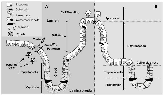
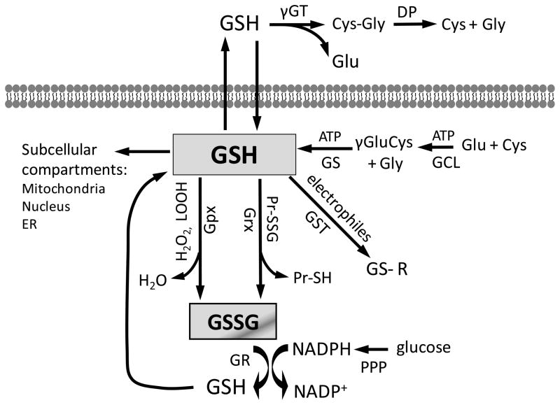
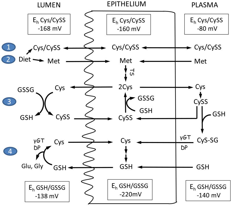
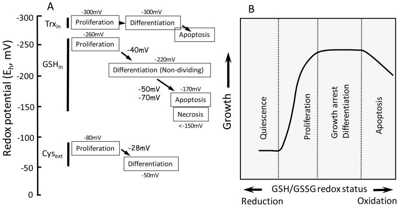
The intestinal tract, known for its capability for self-renew, represents the first barrier of defence between the organism and its luminal environment. The thiol/disulfide redox systems comprising the glutathione/glutathione disulfide (GSH/GSSG), cysteine/cystine (Cys/CySS) and reduced and oxidized thioredoxin (Trx/TrxSS) redox couples play important roles in preserving tissue redox homeostasis, metabolic functions, and cellular integrity. Control of the thiol-disulfide status at the luminal surface is essential for maintaining mucus fluidity and absorption of nutrients, and protection against chemical-induced oxidant injury. Within intestinal cells, these redox couples preserve an environment that supports physiological processes and orchestrates networks of enzymatic reactions against oxidative stress. In this review, we focus on the intestinal redox and antioxidant systems, their subcellular compartmentation, redox signalling and epithelial turnover, and contribution of luminal microbiota, key aspects that are relevant to understanding redox-dependent processes in gut biology with implications for degenerative digestive disorders, such as inflammation and cancer.


Figures

Figure 1
Figure 1. Organization of the small intestinal epithelium
A. The intestinal epithelium, consisting of a single layer of cells, is extensively folded that results in distinct crypt and villus regions. Four types of cells including enterocytes, Paneth, goblet, and enteroendocrine cells reside in the epithelium and are involved in digestive and immunological functions of the intestine. Particulate antigens (pathogens and toxins) are sampled by the M cells present in the gut-associated lymphoid tissues at the luminal site and presented to dendritic cells or other antigen-presenting cells at their basolateral surface. At the basolateral membrane, cells of the intestinal epithelium associate with and are supported by lamina propria. B. The intestinal epithelium is a highly proliferative tissue. Cell proliferation originates at the base of the crypt where intestinal stem cells reside. Progenitors of the stem cells proliferate and migrate bi-directionally. The precursors that migrate toward the tip of the villus differentiate into one of the following cell types: enterocyte, goblet cell, or enteroendocrine cell; the progenitors that migrate toward the bottom of the crypt differentiate into Paneth cells. At 4–5 days post differentiation, villus tip cells die by apoptosis are shedded into the lumen while Paneth cells remain in the crypt for about 23 days and thereafter are phagocytozed.
Figure 2
Figure 2. Cellular GSH homeostasis and GSH-dependent reactions
Intracellular GSH balance is maintained by de novo synthesis, regeneration from GSSG, and extracellular GSH uptake. In transport epithelial cells, such as enterocytes, γ-glutamyl transferase (γ-GT) and dipeptidase (DP) catalyzed the hydrolysis of extracellular GSH to its constituent amino acids, glutamate, cysteine and glycine. Additionally, intestinal epithelial cells can import intact GSH from the lumen via specific plasma membrane transporters. Cytosolic synthesis of GSH takes place in two ATP-dependent reactions catalyzed by glutamate-cysteine ligase (GCL) and glutathione synthase (GS). The intracellular GSH pool, present in millimolar concentrations, is involved in various GSH-dependent reactions. Compartmentation of GSH within the mitochondria, nucleus, or endoplasmic reticulum creates distinct and independently regulated subcellular redox pools. As part of the antioxidant defense system, GSH participates in conjugation reactions catalyzed by glutathione-S-transferases (GSTs), in the reduction of hydrogen peroxide (H2O2) and lipid hydroperoxides (LOOH) catalyzed by glutathione peroxidases (Gpxs), and the reduction of protein-disulfides (PrSSG) catalyzed by glutaredoxins (Grxs). The reduction of glutathione disulfide (GSSG) by glutathione reductase (GR) in the GSH redox cycle regenerates GSH. GSSG reduction occurs at the expense of NADPH, produced from the pentose phosphate pathway (PPP) from glucose oxidation.
Figure 3
Figure 3. Homeostatic control of Cys/CySS redox status in intestinal lumen, intestinal epithelium, and plasma
At the brush-border membrane, uptake of dietary Cys and methionine (1 & 2); Cys/CySS shuttle (3), and γ-glutamyl transferase (γGT) and dipeptidase (DP)-catalyzed hydrolysis of extracellular GSH (4) participate in Cys homeostasis. The Cys/CySS shuttle and luminal GSH hydrolysis maintains the Eh for luminal Cys/CySS at −168mV and that of GSH/GSSG at −138mV. Within the intestinal epithelium, CySS is reduced by GSH, the resultant Cys is exported or utilized in GSH synthesis. Additionally, intracellular Cys is increased through the trans-sulfuration (TS) of imported dietary or circulatory methionine (Met). Plasma Cys exists mainly as CySS, and the redox state of plasma Cys is controlled by thiol/disulfide exchange with liver-derived GSH. The hydrolysis of Cys-GSH mixed disulfide (CyS-SG) releases Cys which is taken into enterocytes by basolateral membrane associated transporters. The Eh for Cys/CySS and GSH/GSSG redox couples are tightly regulated at values of −80mV and −140mV, respectively. Luminal values of Eh for Cys/CySS and GSH/GSSG redox couples were taken from [69] and the plasma values were from [53,106].
Figure 4
Figure 4. Redox potential (Eh) of thiol redox couples during normal phenotypic transition of intestinal cell (A) and cellular responses to changes in the cellular GSH/GSSG redox status (B)
A. Normal intestinal cell transitions from proliferation to differentiation or growth arrest, and apoptosis are associated with increased oxidation of the redox potentials (Eh) of the intracellular GSH/GSSG (GSHin) or extracellular Cys/CySS (Cysext) redox couples. A 40mV oxidation (from −260mV to −220mV) in Eh for cellular GSH/GSSG is associated with cell transition from proliferation to differentiation. An additional 50mV or 70mV oxidation characterizes apoptotic or necrotic cells, respectively [18]. Similarly, an increase in 28mV oxidation of Eh for extracellular Cys/CySS (from −80mV to −50mV) accompanies intestinal cell progression from proliferation to differentiation [99]. In contrast, the Eh for intracellular Trx/TrxSS (Trxin) remains unchanged at −300mV through cell proliferation and differentiation [99], but significant Trx 1 oxidation results in cell apoptosis. B. Fully differentiated intestinal cells typically exhibit a biological constraint to proliferate due to a mitotic block and are arrested in a quiescent state. An imposed change in the status of the cellular GSH/GSSG redox couple during oxidative challenge can initiate entry into cell cycle, differentiation/growth arrest, or apoptosis, depending on the duration and severity of the redox shift.

References

    1. Lipkin M. Proliferation and differentiation of gastrointestinal cells. Physiol Rev. 1973;53:891–915. - PubMed
    1. Cheng H. Origin, differentiation and renewal of the four main epithelial cell types in the mouse small intestine. IV. Paneth cells. Am J Anat. 1974;141:521–535. - PubMed
    1. Spradling A, Drummond-Barbosa D, Kai T. Stem cells find their niche. Nature. 2001;414:98–104. - PubMed
    1. Radtke F, Clevers H. Self-renewal and cancer of the gut: two sides of a coin. Science. 2005;307:1904–1909. - PubMed
    1. Cheng H, Leblond CP. Origin, differentiation and renewal of the four main epithelial cell types in the mouse small intestine. V. Unitarian Theory of the origin of the four epithelial cell types. Am J Anat. 1974;141:537–561. - PubMed

Publication types

LinkOut - more resources