. 2011 Aug 10;13(4):R76.
doi: 10.1186/bcr2924.

Septin 9 isoform expression, localization and epigenetic changes during human and mouse breast cancer progression

Affiliations

Septin 9 isoform expression, localization and epigenetic changes during human and mouse breast cancer progression

Diana Connolly et al. Breast Cancer Res. .

Abstract

Introduction: Altered expression of Septin 9 (SEPT9), a septin coding for multiple isoform variants, has been observed in several carcinomas, including colorectal, head and neck, ovarian and breast, compared to normal tissues. The mechanisms regulating its expression during tumor initiation and progression in vivo and the oncogenic function of its different isoforms remain elusive.

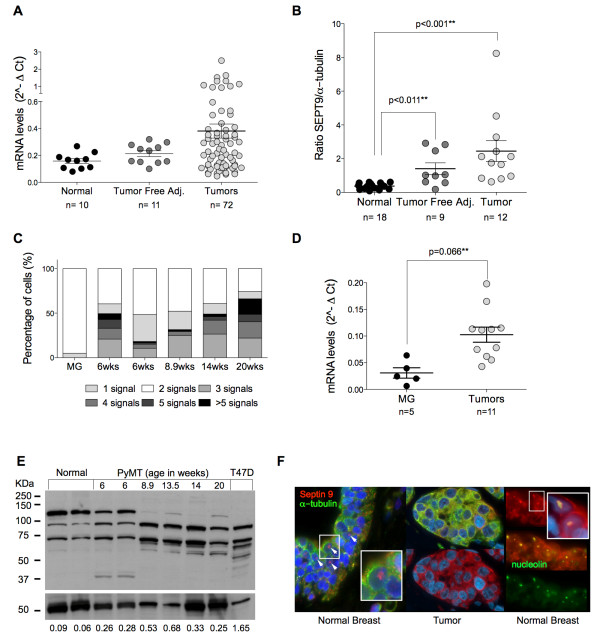
Methods: Using an integrative approach, we investigated SEPT9 at the genetic, epigenetic, mRNA and protein levels in breast cancer. We analyzed a panel of breast cancer cell lines, human primary tumors and corresponding tumor-free areas, normal breast tissues from reduction mammoplasty patients, as well as primary mammary gland adenocarcinomas derived from the polyoma virus middle T antigen, or PyMT, mouse model. MCF7 clones expressing individual GFP-tagged SEPT9 isoforms were used to determine their respective intracellular distributions and effects on cell migration.

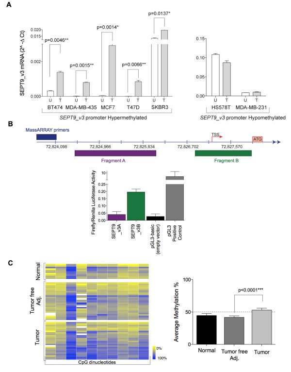
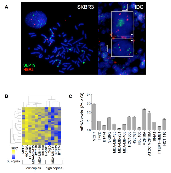
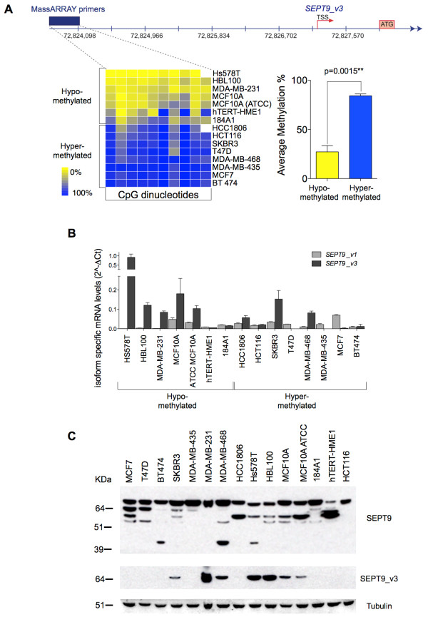
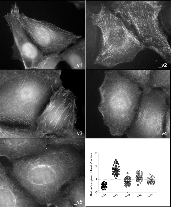
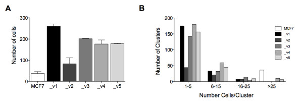
Results: An overall increase in gene amplification and altered expression of SEPT9 were observed during breast tumorigenesis. We identified an intragenic alternative promoter at which methylation regulates SEPT9_v3 expression. Transfection of specific GFP-SEPT9 isoforms in MCF7 cells indicates that these isoforms exhibit differential localization and affect migration rates. Additionally, the loss of an uncharacterized SEPT9 nucleolar localization is observed during tumorigenesis.

Conclusions: In this study, we found conserved in vivo changes of SEPT9 gene amplification and overexpression during human and mouse breast tumorigenesis. We show that DNA methylation is a prominent mechanism responsible for regulating differential SEPT9 isoform expression and that breast tumor samples exhibit distinctive SEPT9 intracellular localization. Together, these findings support the significance of SEPT9 as a promising tool in breast cancer detection and further emphasize the importance of analyzing and targeting SEPT9 isoform-specific expression and function.


Figures

Figure 4
Figure 4
Hypermethylation of the SEPT9_v3 promoter occurs in human breast primary carcinomas. (A) Cell lines were treated with 5-aza-2'-deoxycytidine, and SEPT9_v3 mRNA was quantified. Representative cell lines with hypermethylation (left graph) and hypomethylation (right graph) of the SEPT9_v3 promoter region are displayed. U: untreated cells; T: treated cells. (B) SEPT9_v3 promoter region activity. Top: Location of tested luciferase fragments relative to the DMR. Bottom: Quantification of these fragments via luciferase reporter assay. (C) Supervised clustering of MassARRAY DNA methylation profiles in human primary adenocarcinomas, matching tumor-free areas and normal breast tissues. Right graph: Average levels of DNA methylation between tumors and tumor-free matching samples show a statistically significant difference.
Figure 1
Figure 1
SEPT9 is amplified in human breast cancer. (A) FISH labeling of SEPT9 gene (green) and HER2 gene (red) in SKBR3 breast cancer cells (left panel) and primary invasive ductal carcinoma (IDC) breast tissue (right panel). (B) Unsupervised hierarchical clustering of SEPT9 DNA copy number in human cell lines. Each block represents one cell, and 25 cells were counted for each cell line. (C) Total SEPT9 mRNA levels in the human cell lines (Additional file 1, Supplementary Table 2).
Figure 2
Figure 2
SEPT9 is amplified and overexpressed during breast tumorigenesis. (A) SEPT9 mRNA levels quantified in primary breast tumors (light gray circles), tumor-free adjacent tissues (dark gray) and normal breast tissues from reduction mammoplasty (black circles). (B) Quantification of SEPT9 protein expression in breast primary tumors (light gray circles), tumor-free adjacent tissues (dark gray circles) and normal breast tissues from reduction mammoplasty (black circles). (C) Quantification of Sept9 gene copy number in normal mammary tissues and in primary mammary adenocarcinomas isolated from the PyMT mouse model at various stages of tumorigenesis (MG, normal mammary gland; 6 weeks premalignant hyperplasia, 8.9 weeks adenoma, and 14 and 20 weeks early and late carcinomas, respectively). Legend indicates copy number categories. (D) Sept9 mRNA levels in PyMT murine normal (black circles) and primary adenocarcinoma (light gray circles) mammary tissues. (E) Western blot depicting the pattern of Sept9 isoform (top panel) and α-tubulin (bottom panel) expression in normal mammary tissues and in primary mammary adenocarcinomas isolated from the PyMT mouse model at various stages of tumorigenesis. Values at the bottom indicate Sept9 expression levels normalized to α-tubulin. Only the bands between 37 and 75 kDa were used for quantification. T47D human breast cancer cell line in lane 9 was used as a reference to aid in isoform identification. (F) Left and center panels: immunohistofluorescence labeling of SEPT9 (red) and α-tubulin (green) in normal breast and adenocarcinoma tissues. White arrowheads indicate SEPT9 nuclear staining. Right panel: Colocalization of SEPT9 (red) and nucleolin (green) in normal breast tissues.
Figure 3
Figure 3
Hypermethylation of an alternative promoter in breast tumors downregulates SEPT9_v3. (A) Quantification of DNA methylation at CpG sites upstream of the SEPT9_v3 transcription start site in cell lines targeted by MassARRAY. The unsupervised clustering distributes cell lines into two groups: hypomethylated and hypermethylated (left panel). Differences in the overall methylation percentages between these two groups are shown (right panel). White boxes represent CpG sites that were not analyzable. (B) Quantification of SEPT9_v1 and SEPT9_v3 isoform mRNA levels in human cell lines. (C) Expression of SEPT9 (top panel), SEPT9_v3 isoform (middle panel) and α-tubulin (bottom panel) detected by Western blot in the panel of breast cancer cell lines. Note that with the labeling of recombinant SEPT9_v1 and _v3 by an antibody recognizing both of these isoforms [16], the anti-SEPT9_v3 antibody recognizes the bottom band of the high-molecular-weight isoform doublet.
Figure 5
Figure 5
SEPT9 isoforms localize in different cellular compartments. Fluorescence microscopy of MCF7 clones expressing individual GFP-SEPT9 isoforms (gray scale). Expression of GFP-SEPT9 isoform constructs was quantified and plotted as a ratio of intensities measured outside the region occupied by the nucleus to measurements within the nucleus (bottom right panel). Average ratios located above the dotted line indicate cytoplasmic expression and those below the line indicate nuclear expression.
Figure 6
Figure 6
SEPT9 isoforms alter migratory properties of MCF7. (A) Transwell migration assay comparing MCF7 clones, each expressing a different GFP-SEPT9 isoform fusion construct. Plotting reflects the average number of migrated cells from two biological replicate assays conducted for each isoform. (B) Cell cluster size distribution for each MCF7 clone. The same gray scale is used in (A) and (B) to represent MCF7 and each isoform construct.

Comment in

References

    1. Yamaguchi H, Condeelis J. Regulation of the actin cytoskeleton in cancer cell migration and invasion. Biochim Biophys Acta. 2007;1773:642–652. doi: 10.1016/j.bbamcr.2006.07.001. - DOI - PMC - PubMed
    1. Spiliotis ET, Nelson WJ. Here come the septins: novel polymers that coordinate intracellular functions and organization. J Cell Sci. 2006;119:4–10. doi: 10.1242/jcs.02746. - DOI - PMC - PubMed
    1. Montagna C, Lyu MS, Hunter K, Lukes L, Lowther W, Reppert T, Hissong B, Weaver Z, Ried T. The Septin 9 (MSF) gene is amplified and overexpressed in mouse mammary gland adenocarcinomas and human breast cancer cell lines. Cancer Res. 2003;63:2179–2187. - PubMed
    1. Kuttler F, Mai S. Formation of non-random extrachromosomal elements during development, differentiation and oncogenesis. Semin Cancer Biol. 2007;17:56–64. doi: 10.1016/j.semcancer.2006.10.007. - DOI - PubMed
    1. Sørensen AB, Lund AH, Ethelberg S, Copeland NG, Jenkins NA, Pedersen FS. Sint1, a common integration site in SL3-3-induced T-cell lymphomas, harbors a putative proto-oncogene with homology to the septin gene family. J Virol. 2000;74:2161–2168. doi: 10.1128/JVI.74.5.2161-2168.2000. - DOI - PMC - PubMed

Publication types

MeSH terms