. 2011 Aug 10;30(1):74.
doi: 10.1186/1756-9966-30-74.

Statin-induced apoptosis via the suppression of ERK1/2 and Akt activation by inhibition of the geranylgeranyl-pyrophosphate biosynthesis in glioblastoma

Affiliations

Statin-induced apoptosis via the suppression of ERK1/2 and Akt activation by inhibition of the geranylgeranyl-pyrophosphate biosynthesis in glioblastoma

Masashi Yanae et al. J Exp Clin Cancer Res. .

Abstract

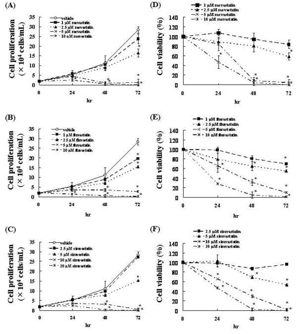
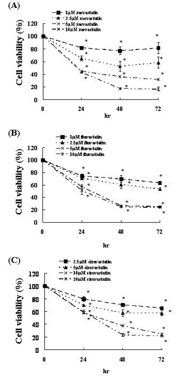
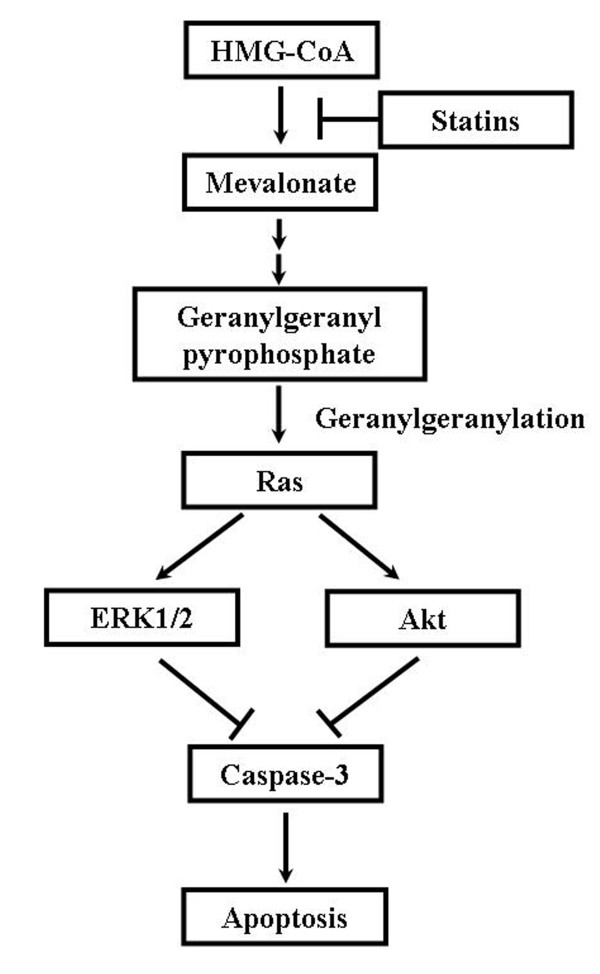
Background: Statins are inhibitors of 3-hydroxy-3-methylglutaryl-coenzyme A reductase, the rate-limiting enzyme in cholesterol synthesis. The inhibition of this key enzyme in the mevalonate pathway leads to suppression of cell proliferation and induction of apoptosis. However, the molecular mechanism of apoptosis induction by statins is not well understood in glioblastoma. In the present study, we attempted to elucidate the mechanism by which statins induce apoptosis in C6 glioma cells.

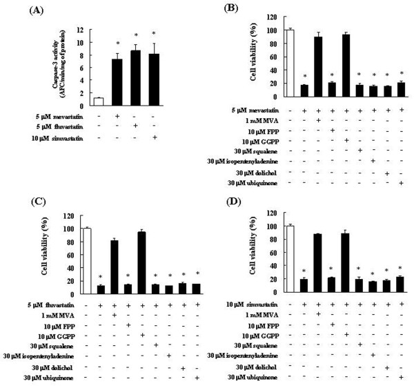
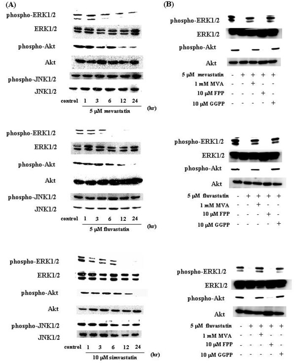
Methods: The cytotoxicity of statins toward the C6 glioma cells were evaluated using a cell viability assay. The enzyme activity of caspase-3 was determined using activity assay kits. The effects of statins on signal transduction molecules were determined by western blot analyses.

Results: We found that statins inhibited cell proliferation and induced apoptosis in these cells. We also observed an increase in caspase-3 activity. The apoptosis induced by statins was not inhibited by the addition of farnesyl pyrophosphate, squalene, ubiquinone, and isopentenyladenine, but by geranylgeranyl-pyrophosphate (GGPP). Furthermore, statins decreased the levels of phosphorylated extracellular signal-regulated kinase 1/2 (ERK1/2) and Akt.

Conclusions: These results suggest that statins induce apoptosis when GGPP biosynthesis is inhibited and consequently decreases the level of phosphorylated ERK1/2 and Akt. The results of this study also indicate that statins could be used as anticancer agents in glioblastoma.


Figures

Figure 1
Figure 1
Effects of statins on C6 glioma cell proliferation and viability. (A-C) C6 glioma cells were incubated at a concentration of 2 × 10⁴ cells/ml for 24 h in a 96-well plate. These cells were treated with various concentrations of statins. After incubation for 24, 48, or 72 h, the number of viable cells was counted by trypan blue staining. The results are representative of 5 independent experiments. *p < 0.01 vs. controls (ANOVA with Dunnett's test). (D-F) C6 glioma cells were treated with various concentrations of statins and trypan blue exclusion test was performed after 24, 48, or 72 h. The results are representative of 5 independent experiments. *p < 0.01 vs. controls (ANOVA with Dunnett's test).
Figure 2
Figure 2
Effects of statins on U251MG cell viability. U251MG cells were treated with various concentrations of statins and trypan blue exclusion test was performed after 24, 48, or 72 h. The results are representative of 5 independent experiments. *p < 0.01 vs. controls (ANOVA with Dunnett's test).
Figure 3
Figure 3
Inhibition of statin-induced apoptosis in C6 glioma cells by intermediates of the mevalonate pathway. (A) Induction of caspase-3-like activity associated with statin-induced cell death. Caspase-3 activity is expressed as pM of proteolytic cleavage of the caspase-3 substrate Asp-Glu-Val-Asp-7-Amino-4-trifluoromethylcoumarin (DEVD-AFC) per h per mg of protein. The results are representative of 5 independent experiments. *p < 0.01 vs. controls (ANOVA with Dunnett's test). (B-D) C6 glioma cells were pretreated with 1 mM mevalonic acid lactone (MVA), 10 μM farnesyl pyrophosphate (FPP), 10 μM geranylgeranyl pyrophosphate (GGPP), 30 μM squalene, 30 μM isopentenyladenine, 30 μM ubiquinone, or 30 μM dolichol for 4 h and then treated with (B) 5 μM mevastatin, (C) 5 μM fluvastatin, or (D) 10 μM simvastatin for 72 h. These results are representative of 5 independent experiments. *p < 0.01 vs. the controls (ANOVA with Dunnett's test).
Figure 4
Figure 4
Statins specifically suppress the activation of Ras/extracellular signal-regulated kinase (ERK) and Ras/Akt pathways in C6 glioma cells. (A) C6 glioma cells were treated with 5 μM mevastatin, 5 μM fluvastatin, or 10 μM simvastatin for 1, 3, 6, 12, or 24 h. Control cells were treated with 0.1% DMSO and cultured in serum-containing medium for 24 h. Whole-cell lysates were generated and immunoblotted with antibodies against phosphorylated ERK1/2 (phospho-ERK1/2), phosphorylated Akt (phospho-Akt), phosphorylated c-Jun N-terminal kinase 1/2 (phospho-JNK1/2), ERK1/2, Akt, and JNK1/2. (B) ERK1/2 and Akt activation in C6 cells to which statins were administered with or without the addition of MVA, FPP, and GGPP. Phospho-ERK1/2, phospho-Akt, ERK1/2, and Akt levels were determined by immunoblotting analysis of the whole-cell lysate.
Figure 5
Figure 5
Schematic representation of interacellular effects of statins in C6 glioma cells.

References

    1. DeAngelis LM. Brain tumors. N Engl J Med. 2001;344:114–123. doi: 10.1056/NEJM200101113440207. - DOI - PubMed
    1. Reardon DA, Wen PY. Therapeutic advances in the treatment of glioblastoma: rationale and potential role of targeted agents. Oncologist. 2006;11:152–164. doi: 10.1634/theoncologist.11-2-152. - DOI - PubMed
    1. Nishida S, Matsuoka H, Tsubaki M, Tanimori Y, Yanae M, Fujii Y, Iwaki M. Mevastatin induces apoptosis in HL60 cells dependently on decrease in phosphorylated ERK. Mol Cell Biochem. 2005;269:109–114. doi: 10.1007/s11010-005-3086-0. - DOI - PubMed
    1. Tsubaki M, Yamazoe Y, Yanae M, Satou T, Itoh T, Kaneko J, Kidera Y, Moriyama K, Nishida S. Blockade of the Ras/MEK/ERK and Ras/PI3K/Akt pathways by statins reduces the expression of bFGF, HGF, and TGF-β as angiogenic factors in mouse osteosarcoma. Cytokine. 2011;54:100–107. doi: 10.1016/j.cyto.2011.01.005. - DOI - PubMed
    1. Wu J, Wong WW, Khosravi F, Minden MD, Penn LZ. Blocking the Raf/MEK/ERK pathway sensitizes acute myelogenous leukemia cells to lovastatin-induced apoptosis. Cancer Res. 2004;64:6461–6468. doi: 10.1158/0008-5472.CAN-04-0866. - DOI - PubMed

Publication types

MeSH terms

Substances