

Evaluation of proximal caries in images resulting from different modes of radiographic digitalization
- PMID: 21831972
- PMCID: PMC3520336
- DOI: 10.1259/dmfr/67185962
Evaluation of proximal caries in images resulting from different modes of radiographic digitalization
Abstract
Objective: The aim of this study was to evaluate the performances of observers in diagnosing proximal caries in digital images obtained from digital bitewing radiographs using two scanners and four digital cameras in Joint Photographic Experts Group (JPEG) and tagged image file format (TIFF) files, and comparing them with the original conventional radiographs.
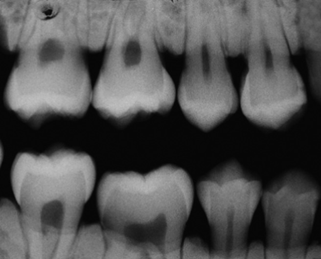
Method: In total, 56 extracted teeth were radiographed with Kodak Insight film (Eastman Kodak, Rochester, NY) in a Kaycor Yoshida X-ray device (Kaycor X-707; Yoshida Dental Manufacturing Co., Tokyo, Japan) operating at 70 kV and 7 mA with an exposure time of 0.40 s. The radiographs were obtained and scanned by CanonScan D646U (Canon USA Inc., Newport News, VA) and Genius ColorPage HR7X (KYE Systems Corp. America, Doral, FL) scanners, and by Canon Powershot G2 (Canon USA Inc.), Canon RebelXT (Canon USA Inc.), Nikon Coolpix 8700 (Nikon Inc., Melville, NY), and Nikon D70s (Nikon Inc.) digital cameras in JPEG and TIFF formats. Three observers evaluated the images. The teeth were then observed under the microscope in polarized light for the verification of the presence and depth of the carious lesions.
Results: The probability of no diagnosis ranged from 1.34% (Insight film) to 52.83% (CanonScan/JPEG). The sensitivity ranged from 0.24 (Canon RebelXT/JPEG) to 0.53 (Insight film), the specificity ranged from 0.93 (Nikon Coolpix/JPEG, Canon Powershot/TIFF, Canon RebelXT/JPEG and TIFF) to 0.97 (CanonScan/TIFF and JPEG) and the accuracy ranged from 0.82 (Canon RebelXT/JPEG) to 0.91 (CanonScan/JPEG).
Conclusion: The carious lesion diagnosis did not change in either of the file formats (JPEG and TIFF) in which the images were saved for any of the equipment used. Only the CanonScan scanner did not have adequate performance in radiography digitalization for caries diagnosis and it is not recommended for this purpose.
Figures
References
-
- Milles DA. The deal of digital: the status of radiographic imaging. Compend Contin Educ Dent 2002;22:1057–1062 - PubMed
-
- Yuasa H, Ariji Y, Ohki M, Naitoh M, Shiojima M, Ushida M, et al. Joint Photographic Experts Group compression of intraoral radiographs for image transmission on the World Wide Web. Oral Surg Oral Med Oral Pathol Oral Radiol Endod 1999;88:93–99 - PubMed
-
- Ohki M, Okano T, Nakamura T. Factors determining the diagnostic accuracy of digitized conventional intraoral radiographs. Dentomaxillofac Radiol 1994;23:77–82 - PubMed
-
- Tosoni GM, Loffredo LCM, Tavano O, Scaf G, Capelozza ALA. Diagnostic quality of conventional and digital radiographic images of dental caries. Rev Odontol UNESP 2001;30:277–290
-
- Attaelmanan A, Borg E, Grondahl HG. Digitisation and display of intra-oral films. Dentomaxillofac Radiol 2000;29:97–102 - PubMed
Publication types
MeSH terms
LinkOut - more resources
Full Text Sources
Medical
