

Comparison of two methods for quantitative assessment of mandibular asymmetry using cone beam computed tomography image volumes
- PMID: 21831974
- PMCID: PMC3277847
- DOI: 10.1259/dmfr/13993523
Comparison of two methods for quantitative assessment of mandibular asymmetry using cone beam computed tomography image volumes
Abstract
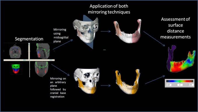
Objectives: The aim of this study was to compare two methods of measuring mandibular asymmetry. The first method uses mirroring of the mandible in the midsagittal plane; the second uses mirroring of the mandible and registration on the cranial base.
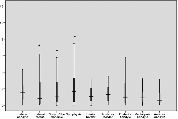
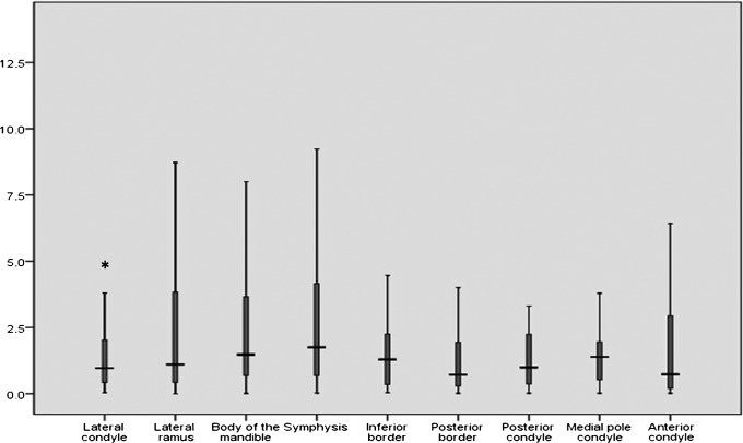
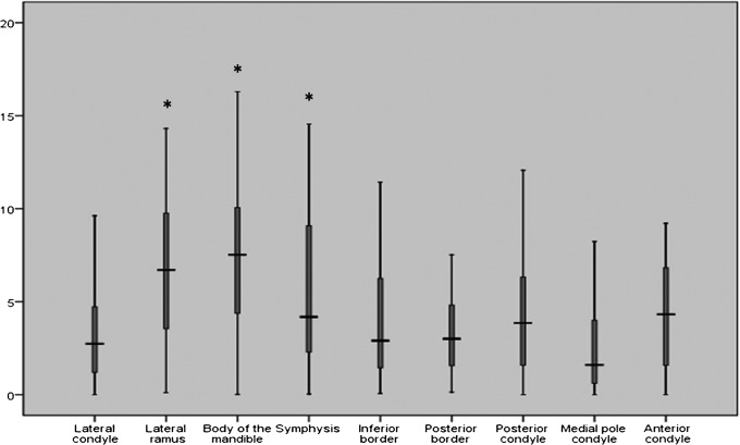
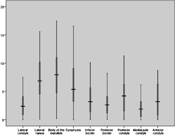
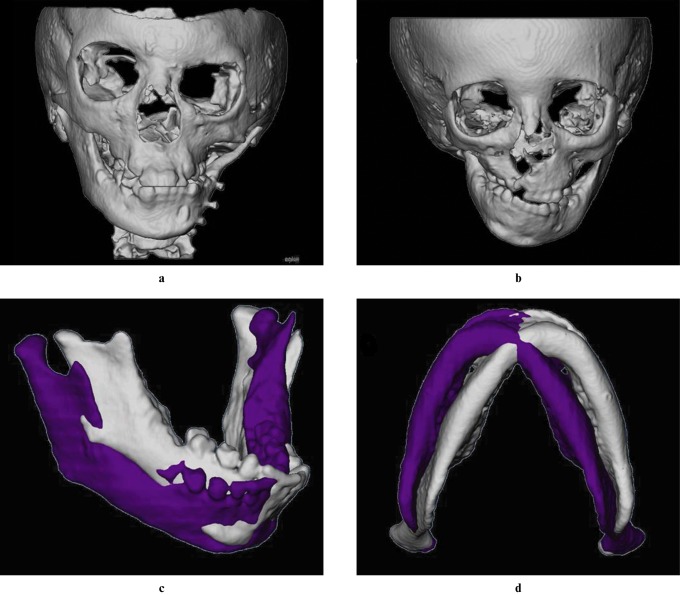
Methods: Surface models were constructed from cone beam CT (CBCT) scans of 50 patients with asymmetry. For the first approach, a midsagittal plane was defined for each patient as the plane passing through nasion, anterior nasal spine and basion. Mirrors for both halves of the mandible were created. The second approach consisted of mirroring the image volume by flipping the left and right sides and then registering the mirrored image onto the cranial base using a mutual information maximization method. Surface distances between hemimandibles and mirrors were calculated for nine regions.
Results: There was no statistically significant difference between the mean surface distance measurements obtained with the two approaches and when comparing both halves in most areas.
Conclusion: Both mirroring techniques provided similar quantification of mandibular asymmetry in this cohort.
Figures
References
-
- Severt TR, Proffit WR. The prevalence of facial asymmetry in the dentofacial deformities population at the University of North Carolina. Int J Adult Orthodon Orthognath Surg 1997;12:171–176 - PubMed
-
- Proffit WR, Fields HW, Jr, Moray LJ. Prevalence of malocclusion and orthodontic treatment need in the United States: Estimates from the NHANES III survey. Int J Adult Orthodon Orthognath Surg 1998;13:97–106 - PubMed
-
- Cevidanes LH, Franco AA, Gerig G, Proffit WR, Slice DE, Enlow DH, et al. Comparison of relative mandibular growth vectors with high-resolution 3-dimensional imaging. Am J Orthod Dentofacial Orthop 2005;128:27–34 - PubMed
