. 2011 Nov;106(5):2471-85.
doi: 10.1152/jn.01097.2010. Epub 2011 Aug 10.

Permanent central synaptic disconnection of proprioceptors after nerve injury and regeneration. II. Loss of functional connectivity with motoneurons

Affiliations

Permanent central synaptic disconnection of proprioceptors after nerve injury and regeneration. II. Loss of functional connectivity with motoneurons

Katie L Bullinger et al. J Neurophysiol. 2011 Nov.

Abstract

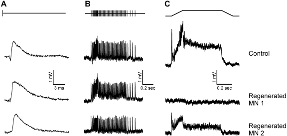
Regeneration of a cut muscle nerve fails to restore the stretch reflex, and the companion paper to this article [Alvarez FJ, Titus-Mitchell HE, Bullinger KL, Kraszpulski M, Nardelli P, Cope TC. J Neurophysiol (August 10, 2011). doi:10.1152/jn.01095.2010] suggests an important central contribution from substantial and persistent disassembly of synapses between regenerated primary afferents and motoneurons. In the present study we tested for physiological correlates of synaptic disruption. Anesthetized adult rats were studied 6 mo or more after a muscle nerve was severed and surgically rejoined. We recorded action potentials (spikes) from individual muscle afferents classified as IA like (*IA) by several criteria and tested for their capacity to produce excitatory postsynaptic potentials (EPSPs) in homonymous motoneurons, using spike-triggered averaging (STA). Nearly every paired recording from a *IA afferent and homonymous motoneuron (93%) produced a STA EPSP in normal rats, but that percentage was only 17% in rats with regenerated nerves. In addition, the number of motoneurons that produced aggregate excitatory stretch synaptic potentials (eSSPs) in response to stretch of the reinnervated muscle was reduced from 100% normally to 60% after nerve regeneration. The decline in functional connectivity was not attributable to synaptic depression, which returned to its normally low level after regeneration. From these findings and those in the companion paper, we put forward a model in which synaptic excitation of motoneurons by muscle stretch is reduced not only by misguided axon regeneration that reconnects afferents to the wrong receptor type but also by retraction of synapses with motoneurons by spindle afferents that successfully reconnect with spindle receptors in the periphery.


Figures

Fig. 1.
Fig. 1.
Response properties distinguishing IA spindle afferents in rat: action potential activity recorded intra-axonally from a medial gastrocnemius (MG) afferent in dorsal root L5 of control rat. A: afferent action potentials (middle trace) pause firing during isometric twitch contraction of MG (top trace) elicited by electrical stimulation of MG nerve. The electrically evoked action potential outlined by rectangle in middle trace shown on expanded time scale in bottom trace displays brief conduction delay of orthodromic action potential from stimulus artifact. B: afferent action potentials (middle trace) firing in response to sequential triangular MG muscle stretches (top trace muscle length, 3 mm amplitude, 750 ms each for ramp stretch and release). Instantaneous firing rates (IFR, bottom trace) from first 2 triangular stretches are superimposed, showing reduced dynamic response in successive stretches (Haftel et al. 2004). C: afferent action potential firing (bottom trace) entrained at 100 Hz (middle trace) by MG muscle vibration (top trace, 80-μm-amplitude oscillation in muscle length 100 ms in duration). D: afferent action potentials (bottom trace) firing in response to ramp-hold-release stretch of MG muscle (top trace muscle length, 3-mm amplitude, 150 ms each for ramp stretch and release, and 0.5-ms hold phase). Afferent IFR (middle trace) with ramp onset (note initial burst firing) and peak and hold phase delineated by upward arrows.
Fig. 2.
Fig. 2.
Nerve regeneration failed to restore normal stretch-evoked synaptic potentials (SSPs): intracellular records collected from MG motoneurons and averaged over multiple trials (10–15) of stretch applied to the MG muscle in anesthetized rats. A: synaptic responses to ramp-hold-release muscle stretch (bottom trace muscle length; 3-mm amplitude, 0.5-s duration) for 1 motoneuron in a control rat and 2 motoneurons (MN1 and MN2) in a rat 13 mo after the MG nerve was cut and surgically rejoined. All control and some regenerated motoneurons (MN1) showed an excitatory SSP (eSSP) consisting of (see arrows in top trace) an initial (i) rapid depolarization, followed by a dynamic (d) response that peaked at the end of the ramp, followed by static (s) depolarization lasting until the end of the hold phase. Many regenerated motoneurons (MN2) failed to show any synaptic response. Responsive (MN1) and unresponsive (MN2) motoneurons could be sampled for the same motor pool in a single regenerated animal. B: cumulative probability plot of SSP amplitudes measured at the peak of the ramp stretch from individual motoneurons pooled for control and regenerated groups. The distribution of SSP amplitudes is left-shifted in regenerated motoneurons compared with controls with 39% of the sample producing no eSSP (12 = 0 mV and 4 < 0 mV). C: synaptic responses to quick stretch of MG muscle (bottom trace muscle length, 1-mm amplitude, 5-ms duration) were also reduced in amplitude (MN1) or absent (MN2). D: the fraction of motoneurons producing excitatory SSPs in control or regenerated groups was similar whether tested with ramp-hold-release stretch or quick stretch.
Fig. 3.
Fig. 3.
Motoneurons recover normal electrical properties whether or not they produce eSSPs after nerve regeneration. Cumulative probability plots for 4 parameters (A–D) from MG motoneurons pooled from control and regenerated groups. Distributions for control vs. regenerated were not significantly different for any parameter (P > 0.5, Student's t-test), and values for motoneurons that failed to produce an eSSP scattered throughout distributions.
Fig. 4.
Fig. 4.
Monosynaptic transmission sustained during physiologically relevant patterns and rates of electrical stimulation. Relationships between peak amplitudes of initial EPSP in train vs. EPSP (A, C) at maximum stimulus frequency (inset in A) or vs. EPSP (B, D) at end of steady-state firing (inset in B) for control (n = 50) and regenerated (n = 21) motoneurons. These measures show little deviation from the initial EPSP: points distribute evenly about the line of identity in A and B, and ratios with EPSPs at maximum and steady-state firing rates approximate 1 (dashed line) in C and D. Independent pooled t-test revealed no significant differences (P > 0.5) between treatment and control groups.
Fig. 5.
Fig. 5.
Synaptic responses to electrical and mechanical stimuli (top traces) compared in selected motoneurons from rats in control and regenerated groups. Columns align responses obtained during MG-nerve electrical stimulation at low frequency (1 pps) (A) and simulated physiological frequencies (see methods) (B) and during MG ramp-hold-release muscle stretch (length, 3 mm amplitude, 150 ms each for ramp stretch and release, and 0.5 ms hold phase) (C). Each of the 3 motoneurons (1 from control rat, MN 1 and MN2 from a single rat 382 days after nerve cut and surgical reunion) responds to electrical stimulation at low or physiological rates, but eSSPs were reduced (MN2) or missing (MN1) in the regenerated motoneuron compared with the normal control.
Fig. 6.
Fig. 6.
Different phases of SSPs were similarly related to each other in controls and regenerated groups. A–C: All 3 permutations of 2-dimensional plots among initial, dynamic, and static phases of eSSP exhibited linear regressions (r) that were not significantly different between control and regenerated groups (P > 0.05, ANCOVA).
Fig. 7.
Fig. 7.
Major reduction in functional synaptic connectivity between afferents that exhibit IA-like (*IA) firing responses and homonymous motoneurons regenerated after nerve section. A: examples of spike-triggered averaged (STA) EPSPs obtained from 3 single IA afferent-motoneuron pairs in the control group (averaged over 267, 1,103, and 609 triggered sweeps of motoneuron membrane potential, respectively, for top, middle, and bottom traces). Dashed line indicates the triggered onset for averaged sweeps. B: all 3 STA EPSPs (averaged over 351, 576, and 834 triggered sweeps for top, middle, and bottom traces, respectively) obtained from a total of 18 single *IA afferent-motoneuron pairs sampled from regenerated group. The STA EPSPs shown in the top and middle rows were produced by the same *IA afferent (conduction delay 1.3 ms) in 2 different motoneurons; the STA EPSP in the bottom row was produced by a different *IA afferent (conduction delay 1.7 ms)-motoneuron pair. C–E: histograms of properties of STA-EPSPs [y-axis shows no. of observations (obs) per bin where each observation was taken from a single IA-motoneuron pair] sampled from control and regenerated groups. C: STA-EPSP peak amplitude, with cases of failure to produce detectable STA-EPSPs entered left of dashed line. STA-EPSPs in the regenerated group were not detected for most (15/18) IA-motoneuron pairs, but the 3 positive cases fell in the normal amplitude range. D: STA-EPSP rise time from base to peak amplitude. E: STA-EPSP onset latency from occurrence of trigger spike in dorsal root. Latencies of the 3 STA-EPSPs from the regenerated group were relatively long and in 2 cases exceeded the normal range.
Fig. 8.
Fig. 8.
Diagrammatic representation of peripheral and central connections made by proprioceptive afferents before nerve cut (normal) and after muscle reinnervation. In A and B the population of muscle spindle and tendon-organ receptors in the MG muscle are modeled, respectively, by 1 coiled and 1 clustered structure contained within the muscle, with 4 sensory neurons variously connecting to the receptors and projecting from the muscle to the spinal cord in which all α-motoneurons (MNs) supplying the same muscle (motor axons not shown) are represented by 3 cells with soma and proximal dendrites located within and distal dendrites extending outside lamina IX (LIX) in the spinal cord. Intramuscular and intraspinal connections are shown for 3 group IA (a, b, c) and 1 group IB (d) afferent before (A) and after (B) reinnervation. In the normal rat (A), the probability that any one IA axon projects into LIX ranges between 0.9 (Collins et al. 1986) and 1.0 (Alvarez et al. 2011), and the probability that each one will synapse (open circles) with any one MN is ∼0.9 (Henneman and Mendell 1981). The product of these probabilities, i.e., the joint probability that afferent b projects to LIX and transmits synaptically with a MN, ranges from 0.8 to 0.9. Arrows on afferents a and c point to their widespread connections with MNs; the IB afferent d connected with the tendon organ does not project into LIX or connect with MNs. After nerve cut and regeneration (B), many IA synapses (VGLUT1) are lost in LIX on MN soma (∼85% loss) and proximal dendrites (∼50% loss). Afferent a fails to reconnect with a muscle spindle and does not respond to muscle stretch but maintains functional synapses that can be activated by electrical stimulation to produce EPSPs in all MNs, including the leftmost MN, which has lost input from all stretch-activated afferents. Afferents b, c, and d reinnervate the spindle receptor and are designated *IA because they exhibit IA-like firing in response to muscle stretch. Afferent b represents the independent probabilities (see text) that 1) an individual *IA sends a projection into LIX (P = 0.2–0.4) and 2) that an *IA that does project to LIX transmits excitation (P = 0.4–0.7). The joint probability that afferent b projects to LIX and transmits synaptically with a MN ranges from 0.08 to 0.28. Afferents c and d represent *IAs that do not project to LIX, which might occur because an original IA afferent c retracts its projection into LIX (double arrowhead) and loses connection with MNs or because an original IB afferent d, which has no monosynaptic connections with MNs, inappropriately reinnervates a muscle spindle receptor.

Similar articles

Cited by

References

    1. Abelew TA, Miller MD, Cope TC, Nichols TR. Local loss of proprioception results in disruption of interjoint coordination during locomotion in the cat. J Neurophysiol 84: 2709–2714, 2000 - PubMed
    1. Alvarez FJ, Bullinger KL, Titus HE, Nardelli P, Cope TC. Permanent reorganization of Ia afferent synapses on motoneurons after peripheral nerve injuries. Ann NY Acad Sci 1198: 231–241, 2010 - PMC - PubMed
    1. Alvarez FJ, Nardelli P, Bullinger KL, Ukpabi N, Crum JM, Zerda R, Kraszpulski M, Cope TC. VGLUT1 content in central synapses of normal and regenerated Ia afferents (Abstract). 2008 Neuroscience Meeting Planner Washington, DC: Society for Neuroscience, 2008, Program No. 74.1 (online)
    1. Alvarez FJ, Titus-Mitchell HE, Bullinger KL, Kraszpulski M, Nardelli P, Cope TC. Permanent central synaptic disconnection of proprioceptors after nerve injury and regeneration. I. Loss of VGLUT1/IA synapses on motoneurons. J Neurophysiol (August 10, 2011). doi:10.1152/jn.01095.2010 - DOI - PMC - PubMed
    1. Banks RW, Barker D. Neuronal specificities in the reinnervation of muscle spindles. In: Plasticity of Motoneuronal Connections, edited by Wering A. Amsterdam: Elsevier, 1991, p. 241–249

Publication types

MeSH terms