. 2011 Oct 7;286(40):35187-95.
doi: 10.1074/jbc.M111.250746. Epub 2011 Aug 8.

Interaction studies between the chloroplast signal recognition particle subunit cpSRP43 and the full-length translocase Alb3 reveal a membrane-embedded binding region in Alb3 protein

Affiliations

Interaction studies between the chloroplast signal recognition particle subunit cpSRP43 and the full-length translocase Alb3 reveal a membrane-embedded binding region in Alb3 protein

Beatrix Dünschede et al. J Biol Chem. .

Abstract

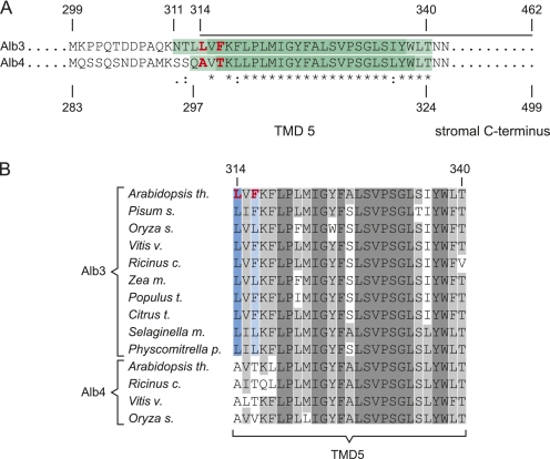
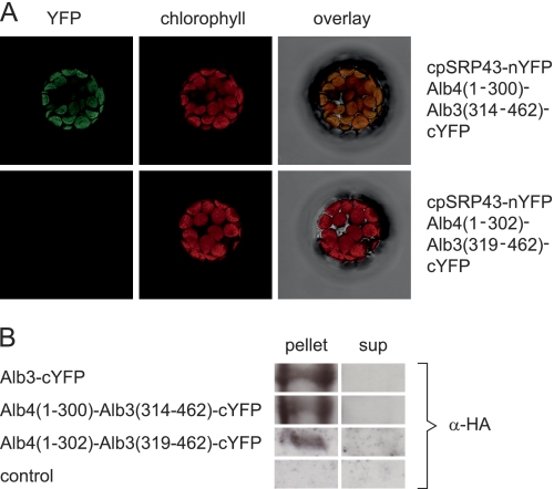
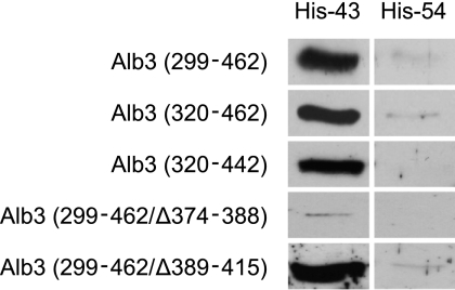
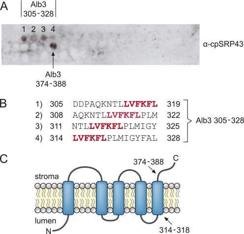
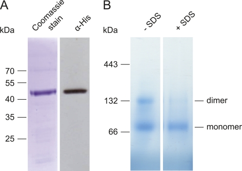
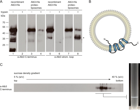
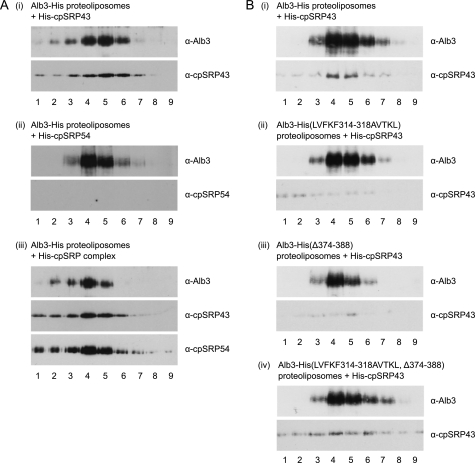
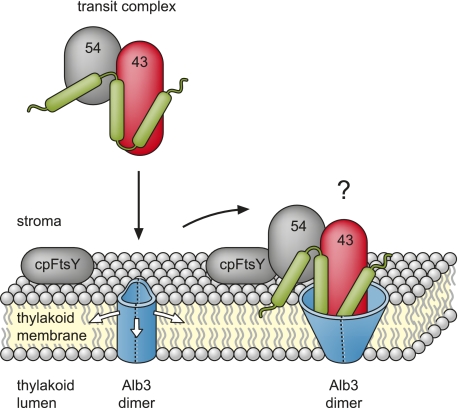
Posttranslational targeting of the light-harvesting chlorophyll a,b-binding proteins depends on the function of the chloroplast signal recognition particle, its receptor cpFtsY, and the translocase Alb3. The thylakoid membrane protein Alb3 of Arabidopsis chloroplasts belongs to the evolutionarily conserved YidC/Oxa1/Alb3 protein family; the members of this family facilitate the insertion, folding, and assembly of membrane proteins in bacteria, mitochondria, and chloroplasts. Here, we analyzed the interaction sites of full-length Alb3 with the cpSRP pathway component cpSRP43 by using in vitro and in vivo studies. Bimolecular fluorescence complementation and Alb3 proteoliposome studies showed that the interaction of cpSRP43 is dependent on a binding domain in the C terminus of Alb3 as well as an additional membrane-embedded binding site in the fifth transmembrane domain (TMD5) of Alb3. The C-terminal binding domain was mapped to residues 374-388, and the binding domain within TMD5 was mapped to residues 314-318 located close to the luminal end of TMD5. A direct binding between cpSRP43 and these binding motifs was shown by pepspot analysis. Further studies using blue-native gel electrophoresis revealed that full-length Alb3 is able to form dimers. This finding and the identification of a membrane-embedded cpSRP43 binding site in Alb3 support a model in which cpSRP43 inserts into a dimeric Alb3 translocation pore during cpSRP-dependent delivery of light-harvesting chlorophyll a,b-binding proteins.


Figures

FIGURE 1.
FIGURE 1.
Prediction and alignment of TMD5 of Alb3 and Alb4 from A. thaliana. A, the regions of TMD5 from Alb3 and Alb4 were predicted by ARAMEMNON. Sequence alignment was performed using ClustalW2. The green region represents the consensus prediction of the different programs. The maximal predicted region of the transmembrane domains is indicated by light green boxes. The region of Alb3 required for cpSRP43 binding in vivo (residues 314–462) (24) is shown by a black line. Nonconservative amino acid exchanges in TMD5 within this binding region are marked in red. The positions of the amino acids of Alb3 and Alb4 are given by numbers. Symbols display the degree of conservation: identical residues (star), conserved substitution (colon), semiconserved substitution (dot). B, TMD5 of Alb3 and Alb4 from various organisms are compared. Amino acid Leu-314 (colored in blue) is invariant, and Phe-316 (colored in light blue) is highly conserved among the Alb3 proteins, but both are absent in Alb4. Invariant and highly conserved residues present in both Alb3 and Alb4 are boxed in dark and light gray, respectively. These residues are not specifically conserved in either Alb3 or Alb4. Numbering of the residues corresponds to the A. thaliana Alb3 sequence.
FIGURE 2.
FIGURE 2.
Analysis of the interaction between cpSRP43 and Alb4/Alb3 fusion constructs using BiFC. A, Arabidopsis mesophyll protoplasts were transiently transformed with two plasmids encoding cpSRP43-nYFP and the Alb4/Alb3-cYFP fusion constructs Alb4(1–300)Alb3(314–462) and Alb4(1–302)Alb3(319–462). All control reactions using a combination of the indicated constructs and control plasmids encoding a chloroplast transit sequence fused to nYFP or cYFP were negative (data not shown). B, protoplasts transfected with Alb3-cYFP, Alb4(1–300)Alb3(314–462), and Alb4(1–302)Alb3(319–462) were lysed and centrifuged, and the supernatant (sup) was precipitated with TCA. The membrane pellet was washed with 0.2 m NaOH and centrifuged. Western blot analysis with the pellet and the precipitate of the supernatant was done using an α-HA antibody. For the control, untransfected protoplasts were used.
FIGURE 3.
FIGURE 3.
The C-terminal region of Alb3 is sufficient for cpSRP43 binding in vitro. In vitro pulldown assays were performed using recombinant His-cpSRP43 (His-43) and the indicated in vitro translation products of Alb3. Control reactions were performed with recombinant His-cpSRP54 (His-54) instead of His-cpSRP43. His-tagged proteins were repurified by Ni-NTA resin, and coeluted Alb3 proteins were detected by immunoblotting using an antibody against Alb3 (for details, see “Experimental Procedures”).
FIGURE 4.
FIGURE 4.
Alb3 pepscan analysis reveals two binding sites for recombinant cpSRP43. A, the interaction of His-cpSRP43 and Alb3 (residues 299–462) was analyzed using a pepscan approach. Recombinant His-cpSRP43 was incubated in a final concentration of 5 μg/ml with a peptide library comprising the Alb3 amino acids 299–462. The peptide library contained 51 15-mer peptides, overlapping by 12 residues. Bound protein was detected by immunoblotting using an antibody against cpSRP43. Detected spots of bound recombinant cpSRP43 correspond to residues 305–328 (spots 1–4) and 374–388 of Alb3. B, amino acid sequences of the 15-mer peptides corresponding to spots 1–4 (residues 305–328) of Alb3 are shown. The LVFKFL motif is present in each of the four peptides and marked in red. C, schematic representation of Alb3 topology. The cpSRP43 binding sites in the stromal C terminus and the membrane-embedded region of Alb3 are indicated.
FIGURE 5.
FIGURE 5.
SDS- and BN-PAGE analysis of recombinant Alb3-His. A, Alb3-His was expressed in E. coli and purified using Ni-NTA agarose beads as described in detail under “Experimental Procedures. ” The eluate was analyzed by SDS-PAGE and Coomassie staining or Western blotting using an α-His antibody. B, Alb3-His oligomerization was analyzed by BN-PAGE according to the manufacturer's instructions (NativePAGETM Novex® Bis-Tris Gel System; Invitrogen). Alb3-His could be detected as monomer and dimer. The signal of the Alb3-His dimer disappeared after denaturation with 5% (w/v) SDS. The higher apparent molecular mass of monomeric Alb3-His in BN-PAGE compared with SDS-PAGE is probably due to suboptimal binding of Coomassie dye causing an incomplete charge shift. This running behavior was described for several integral membrane proteins (44).
FIGURE 6.
FIGURE 6.
Generation of Alb3-His proteoliposomes. A, trypsin digestion experiments were performed to analyze the Alb3-His orientation in proteoliposomes. Samples equal to 250 ng of recombinant Alb3-His protein (lanes 1, 2, 5, and 6) and Alb3-His integrated into liposomes (lanes 3, 4, 7, and 8) were analyzed by SDS-PAGE and Western blotting. A reconstitution efficiency of 30–50% was calculated. Recombinant Alb3-His samples contained some unspecific degradation products (lanes 1 and 5) that were not integrated into the liposomes (lanes 3 and 7). The trypsin treatment resulted in a complete digestion of recombinant Alb3-His (lanes 2 and 6). The trypsin treatment of proteoliposomes (lanes 4 and 8) generated a specific product of ∼35 kDa (lane 8, marked with an asterisk), that could only be detected with the antibody directed against the first stromal loop region of Alb3 (lane 8) and not with the antibody directed against the C terminus (lane 4). Trypsin-digested proteoliposome samples shown in lanes 4 and 8 contained some residual full-length Alb3-His. B, schematic overview shows the orientation of Alb3 with its C terminus exposed to the exterior and its N terminus located in the interior of the liposomes. C, Alb3-His proteoliposomes were loaded onto the top of sucrose gradients and centrifuged. Fractions were analyzed by SDS-PAGE and Western blotting. Alb3-His could be detected in the fractions corresponding to the visible band of proteoliposomes in the sucrose gradients, which have a sucrose density of ∼30% (w/v).
FIGURE 7.
FIGURE 7.
Binding of cpSRP43, cpSRP54, or the preformed cpSRP complex to Alb3-His proteoliposomes and cpSRP43 binding to mutated Alb3-His variants integrated into proteoliposomes. A, Alb3-His proteoliposomes were generated as described in detail under “Experimental Procedures ” and incubated with recombinant His-cpSRP43 (i), His-cpSRP54 (ii), or a His-cpSRP complex (iii), loaded on top of sucrose density gradients, and centrifuged. Fractions were analyzed by SDS-PAGE and Western blotting, and samples corresponding to the visible liposome band in the sucrose gradients (see also Fig. 6) are displayed. Unbound protein was detected in the upper fractions of the gradients (data not shown), whereas the bound protein comigrated with the different Alb3-His proteoliposomes into the sucrose density gradient. B, proteoliposomes containing Alb3-His (i), Alb3-His(LVFKF314–318AVTKL) (ii), Alb3-His(Δ374–388) (iii), and Alb3-His(LVFKF314–318AVTKL, Δ374–388) (iv) were incubated with recombinant His-cpSRP43 and analyzed as described in A.
FIGURE 8.
FIGURE 8.
Model of transit complex docking to a dimeric Alb3 insertion pore in the thylakoid membrane. The model suggests that dimeric Alb3 forms a translocation pore with stroma-exposed C termini. The transit complex docks to the translocase in a cpSRP43-mediated interaction. The cpSRP43 binding sites are located in the stroma-exposed C terminus as well as in TMD5. Therefore, cpSRP43 is postulated to immerge into the Alb3 pore. A coinsertion of the LHCP can be hypothesized because a binding region for LHCP has been localized to a region involving TMD5 (24). The precise functions of cpSRP54 and cpFtsY in the docking process remain to be clarified.

Similar articles

Cited by

References

    1. Kuhn A., Stuart R., Henry R., Dalbey R. E. (2003) Trends Cell Biol. 13, 510–516 - PubMed
    1. Luirink J., Samuelsson T., de Gier J. W. (2001) FEBS Lett. 501, 1–5 - PubMed
    1. Wang P., Dalbey R. E. (2011) Biochim. Biophys. Acta 1808, 866–875 - PubMed
    1. du Plessis D. J., Nouwen N., Driessen A. J. (2011) Biochim. Biophys. Acta 1808, 851–865 - PubMed
    1. Nouwen N., Driessen A. J. (2002) Mol. Microbiol. 44, 1397–1405 - PubMed

Publication types