

Alteration of developmental and pathological retinal angiogenesis in angptl4-deficient mice
- PMID: 21832056
- PMCID: PMC3196087
- DOI: 10.1074/jbc.M111.220061
Alteration of developmental and pathological retinal angiogenesis in angptl4-deficient mice
Abstract
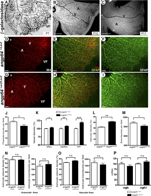
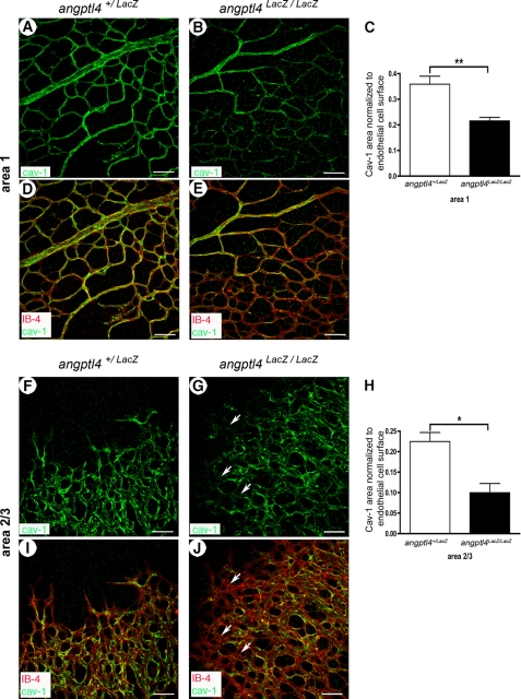
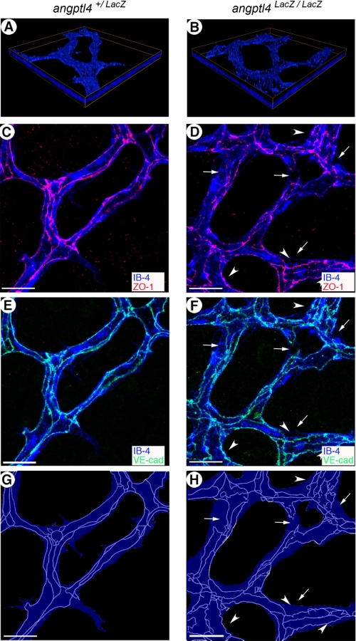
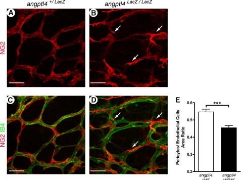
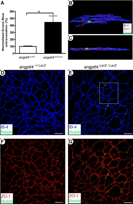
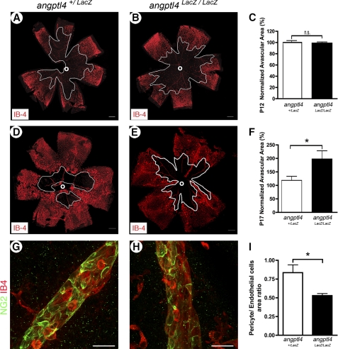
Proper vessel maturation, remodeling of endothelial junctions, and recruitment of perivascular cells is crucial for establishing and maintaining vessel functions. In proliferative retinopathies, hypoxia-induced angiogenesis is associated with disruption of the vascular barrier, edema, and vision loss. Therefore, identifying factors that regulate vascular maturation is critical to target pathological angiogenesis. Given the conflicting role of angiopoietin-like-4 (ANGPTL4) reported in the current literature using gain of function systems both in vitro and in vivo, the goal of this study was to characterize angiogenesis, focusing on perinatal retinal vascularization and pathological circumstances in angpl4-deficient mice. We report altered organization of endothelial junctions and pericyte coverage, both leading to impaired angiogenesis and increased vascular leakage that were eventually caught up, suggesting a delay in vessel maturation. In a model of oxygen-induced retinopathy, pathological neovascularization, which results from tissue hypoxia, was also strongly inhibited in angptl4-deficient mice. This study therefore shows that ANGPTL4 tunes endothelial cell junction organization and pericyte coverage and controls vascular permeability and angiogenesis, both during development and in pathological conditions.
Figures
Similar articles
-
Natriuretic Peptides Attenuate Retinal Pathological Neovascularization Via Cyclic Guanosine Monophosphate Signaling in Pericytes and Astrocytes.Arterioscler Thromb Vasc Biol. 2020 Jan;40(1):159-174. doi: 10.1161/ATVBAHA.119.313400. Epub 2019 Oct 17. Arterioscler Thromb Vasc Biol. 2020. PMID: 31619060
-
ANGPTL4-αvβ3 interaction counteracts hypoxia-induced vascular permeability by modulating Src signalling downstream of vascular endothelial growth factor receptor 2.J Pathol. 2016 Dec;240(4):461-471. doi: 10.1002/path.4805. Epub 2016 Oct 21. J Pathol. 2016. PMID: 27577973
-
Hypoxic retinal Muller cells promote vascular permeability by HIF-1-dependent up-regulation of angiopoietin-like 4.Proc Natl Acad Sci U S A. 2013 Sep 3;110(36):E3425-34. doi: 10.1073/pnas.1217091110. Epub 2013 Aug 19. Proc Natl Acad Sci U S A. 2013. PMID: 23959876 Free PMC article.
-
Role of Angptl4 in vascular permeability and inflammation.Inflamm Res. 2014 Jan;63(1):13-22. doi: 10.1007/s00011-013-0678-0. Epub 2013 Oct 31. Inflamm Res. 2014. PMID: 24173241 Review.
-
Tie-1: A potential target for anti-angiogenesis therapy.J Huazhong Univ Sci Technolog Med Sci. 2015 Oct;35(5):615-622. doi: 10.1007/s11596-015-1479-1. Epub 2015 Oct 22. J Huazhong Univ Sci Technolog Med Sci. 2015. PMID: 26489611 Review.
Cited by
-
Angiopoietin-Like Proteins in Angiogenesis, Inflammation and Cancer.Int J Mol Sci. 2018 Feb 1;19(2):431. doi: 10.3390/ijms19020431. Int J Mol Sci. 2018. PMID: 29389861 Free PMC article. Review.
-
A review of the multifunctionality of angiopoietin-like 4 in eye disease.Biosci Rep. 2018 Sep 13;38(5):BSR20180557. doi: 10.1042/BSR20180557. Print 2018 Oct 31. Biosci Rep. 2018. PMID: 30049845 Free PMC article. Review.
-
Suppression of angiopoietin-like 4 reprograms endothelial cell metabolism and inhibits angiogenesis.Nat Commun. 2023 Dec 12;14(1):8251. doi: 10.1038/s41467-023-43900-0. Nat Commun. 2023. PMID: 38086791 Free PMC article.
-
Thrombolytic tPA-induced hemorrhagic transformation of ischemic stroke is mediated by PKCβ phosphorylation of occludin.Blood. 2022 Jul 28;140(4):388-400. doi: 10.1182/blood.2021014958. Blood. 2022. PMID: 35576527 Free PMC article.
-
c-Myc is essential to prevent endothelial pro-inflammatory senescent phenotype.PLoS One. 2013 Sep 6;8(9):e73146. doi: 10.1371/journal.pone.0073146. eCollection 2013. PLoS One. 2013. PMID: 24039874 Free PMC article.
References
-
- Holderfield M. T., Hughes C. C. (2008) Circ. Res. 102, 637–652 - PubMed
-
- Germain S., Monnot C., Muller L., Eichmann A. (2010) Curr. Opin. Hematol. 17, 245–251 - PubMed
-
- Adams R. H., Alitalo K. (2007) Nat. Rev. Mol. Cell Biol. 8, 464–478 - PubMed
-
- Phng L. K., Gerhardt H. (2009) Dev. Cell 16, 196–208 - PubMed
Publication types
MeSH terms
Substances
LinkOut - more resources
Full Text Sources
Other Literature Sources
Molecular Biology Databases
