. 2011 Sep 30;286(39):34448-56.
doi: 10.1074/jbc.M111.273342. Epub 2011 Aug 10.

Plasma membrane association of p63 Rho guanine nucleotide exchange factor (p63RhoGEF) is mediated by palmitoylation and is required for basal activity in cells

Affiliations

Plasma membrane association of p63 Rho guanine nucleotide exchange factor (p63RhoGEF) is mediated by palmitoylation and is required for basal activity in cells

Mohamed Aittaleb et al. J Biol Chem. .

Abstract

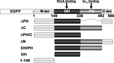
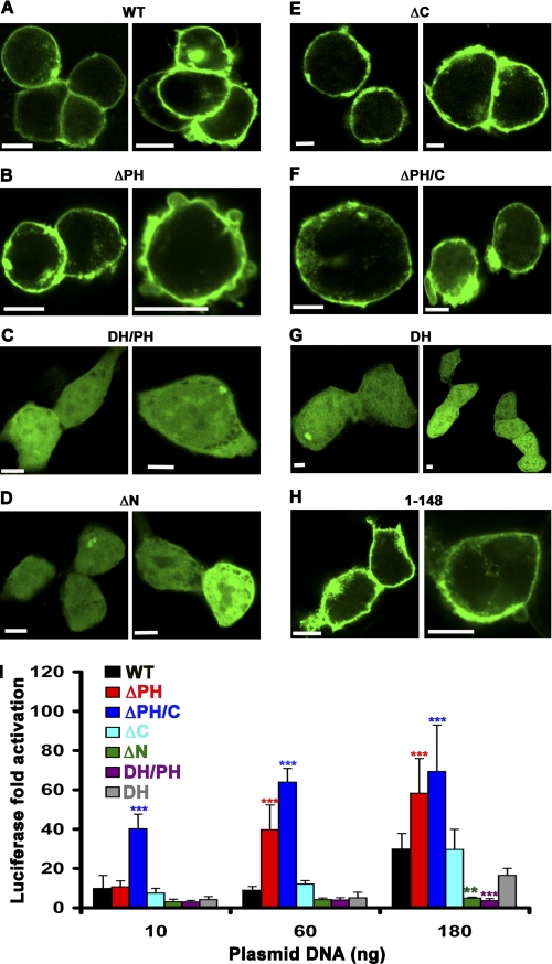
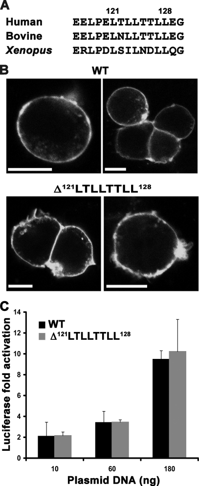
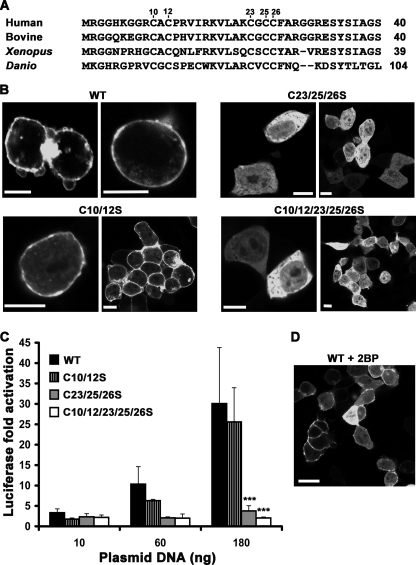
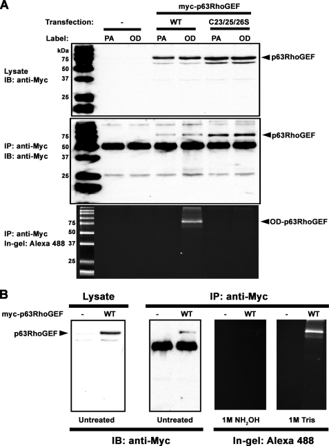
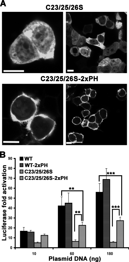
Activation of G protein-coupled receptors at the cell surface leads to the activation or inhibition of intracellular effector enzymes, which include various Rho guanine nucleotide exchange factors (RhoGEFs). RhoGEFs activate small molecular weight GTPases at the plasma membrane (PM). Many of the known G protein-coupled receptor-regulated RhoGEFs are found in the cytoplasm of unstimulated cells, and PM recruitment is a critical aspect of their regulation. In contrast, p63RhoGEF, a Gα(q)-regulated RhoGEF, appears to be constitutively localized to the PM. The objective of this study was to determine the molecular basis for the localization of p63RhoGEF and the impact of its subcellular localization on its regulation by Gα(q). Herein, we show that the pleckstrin homology domain of p63RhoGEF is not involved in its PM targeting. Instead, a conserved string of cysteines (Cys-23/25/26) at the N terminus of the enzyme is palmitoylated and required for membrane localization and full basal activity in cells. Conversion of these residues to serine relocates p63RhoGEF from the PM to the cytoplasm, diminishes its basal activity, and eliminates palmitoylation. The activity of palmitoylation-deficient p63RhoGEF can be rescued by targeting to the PM by fusion with tandem phospholipase C-δ1 pleckstrin homology domains or by co-expression with wild-type Gα(q) but not with palmitoylation-deficient Gα(q). Our data suggest that p63RhoGEF is regulated chiefly through allosteric control by Gα(q), as opposed to other known Gα-regulated RhoGEFs, which are instead sequestered in the cytoplasm, perhaps because of their high basal activity.


Figures

FIGURE 1.
FIGURE 1.
Schematic representation of EGFP-p63RhoGEF variants used to test subcellular localization of p63RhoGEF and its ability to activate RhoA in HEK293 cells. p63RhoGEF is composed of central tandem DH and PH domains, bracketed by N-terminal (N-ter) and C-terminal (C-ter) extensions with relatively low sequence complexity. The DH domain is the catalytic center of the enzyme and represents the primary RhoA binding site, whereas the PH domain is the primary binding site of Gαq. For visualization with confocal microscopy, EGFP was fused to the N terminus of each p63RhoGEF variant (shown only for wild-type p63RhoGEF).
FIGURE 2.
FIGURE 2.
The N-terminal extension of p63RhoGEF is necessary and sufficient for PM association and for high basal activity in cells. A–H, confocal microscopy images of HEK293 cells transfected with 250 ng of DNA encoding the indicated EGFP-p63RhoGEF variant. A, wild-type p63RhoGEF (WT); B, ΔPH; C, DH/PH; D, ΔN; E, ΔC; F, ΔPH/C; G, DH; H, 1–148. Only variants that contain the N-terminal 148 amino acids of p63RhoGEF are localized to the PM. Data shown are representative of 3–5 independent experiments. Scale bar, 10 μm. I, membrane localization correlates with high basal SRE activity in HEK293 cells. EGFP-p63RhoGEF-induced SRE activity is reported as the -fold increase of normalized luciferase over that of EGFP alone as a function of the increasing amount of transfecting EGFP-p63RhoGEF plasmid. ΔN and DH/PH, which are not PM-associated, have negligible activity. The DH domain, which is localized to the cytoplasm, shows increased activity compared with DH/PH due to the absence of autoinhibition imposed by the PH domain but still exhibits lower activity than wild-type p63RhoGEF. The highest activities are obtained for ΔPH and ΔPH/C, presumably due to loss of PH domain-mediated inhibition combined with retention of membrane localization. The data shown are representative of four independent experiments with each data point measured in triplicate and reported as the mean ± S.D. (error bars). Asterisks indicate a significant difference from wild type (**, p < 0.01; ***, p < 0.001).
FIGURE 3.
FIGURE 3.
A conserved hydrophobic di-Leu-based motif in the N terminus of p63RhoGEF is not required for PM association or for SRE activity in cells. A, amino acid sequence alignment showing conservation of the di-Leu-based hydrophobic motif in human (Q86VW2), bovine (XP_002687606.1), and Xenopus (NP_001123746.1) orthologs. B, deletion of the 121LTLLTTLL128 motif does not disrupt PM localization of EGFP-p63RhoGEF. Data shown are representative of four experiments. Scale bar, 10 μm. C, the Δ121LTLLTTLL128 variant also retains wild-type basal activity in cells. The data shown are representative of five independent experiments performed in triplicate. Error bars, S.D.
FIGURE 4.
FIGURE 4.
A conserved N-terminal Cys string is required for PM association and optimal basal activity. A, alignment of the N-terminal region of p63RhoGEF orthologs reveals two conserved Cys strings. Accession numbers are Q86VW2 (human), XP_002687606.1 (bovine), NP_001123746.1 (Xenopus), and XP_698049.4 (Danio). B, subcellular distribution of EGFP-p63RhoGEF Cys string mutants. The C23S/C25S/C26S mutant (C23/25/26S) is found in the cytoplasm, whereas the C10S/C12S mutant (C10/12S) is retained at the PM. Mutation of both Cys strings to Ser also confers cytoplasmic localization. Data shown are representative of four experiments. Scale bar, 10 μm. C, SRE activities are abolished for the C23S/C25S/C26S and C10S/C12S/C23S/C25S/C26S mutants and reduced for the C10S/C12S mutant of p63RhoGEF compared with wild type. The data shown are representative of 3–5 independent experiments performed in triplicate. ***, significant difference at the p < 0.001 level compared with wild type. D, 2BP partially inhibits membrane targeting of p63RhoGEF. Confocal microscopy shows that about 50% of 2BP-treated cells exhibit cytoplasmic distribution of wild-type EGFP-p63RhoGEF. Data shown are representative of two experiments. Scale bar, 20 μm. Error bars, S.D.
FIGURE 5.
FIGURE 5.
p63RhoGEF is palmitoylated at Cys-23/25/26 in cells. A, HEK293 cells transfected with either wild-type (WT), C23S/C25S/C26S (C23/25/26S), or no (control) Myc-p63RhoGEF were incubated in medium containing 100 μm PA or 100 μm 17-ODYA (OD) for 6 h. Shown are immunoblots (IB) of lysates (top) and immunoprecipitates (IP) (middle) using an anti-Myc antibody showing expression and pull-down of WT and C23S/C25S/C26S, respectively. In-gel fluorescence detection of Alexa-488 in immunoprecipitated samples (bottom) shows a fluorescent band only for wild-type p63RhoGEF when labeled with OD (OD-p63RhoGEF). The data shown are representative of three independent experiments. B, confirmation of thioester linkage at Cys-23/25/26. HEK293 cells expressing Myc-p63RhoGEF were incubated in medium containing 100 μm 17-ODYA for 6 h. Lysates were immunoprecipitated with anti-Myc antibody, and 17-ODYA-labeled p63RhoGEF was detected with Alexa488-azide using click chemistry. Samples were resolved by SDS-PAGE on triplicate gels. One gel was soaked in 1 m Tris (pH 7.5), and another was soaked in 1 m NH2OH (pH 7.5) for 16 h before detection of in-gel fluorescence. The third gel was transferred to a nitrocellulose membrane for immunoblotting. The fluorescent band corresponding to 17-ODYA-labeled p63RhoGEF is eliminated upon treatment with 1 m NH2OH, pH 7.5, but unaffected by treatment with 1 m Tris, pH 7.5. The data shown are representative of two experiments.
FIGURE 6.
FIGURE 6.
Targeting palm p63RhoGEF to the PM rescues its activity. A, confocal images of HEK293 cells showing PM translocation of palm EGFP-p63RhoGEF (C23/25/26S) when fused to two tandem PH domains from PLCδ1 (C23/25/26S-2xPH). Data shown are representative of three experiments. Scale bar, 10 μm. B, SRE activities of wild-type and palm EGFP-p63RhoGEF in the absence (WT and C23/25/26S, respectively) or presence (WT-2xPH and C23/25/26S-2xPH, respectively) of the two tandem PH domains from PLCδ1. The data shown are representative of four independent experiments, each performed in triplicate. Significance between the indicated columns is indicated by asterisks (**, p < 0.01; ***, p < 0.001). Error bars, S.D.
FIGURE 7.
FIGURE 7.
q rescues the membrane localization and activity of palm p63RhoGEF in a palmitoylation-dependent manner. A, confocal images of HEK293 cells co-transfected with EGFP-p63RhoGEF and mRFP-Gαq. Wild-type (WT) Gαq, but not Gαq-C9S/C10S (C9/10S), recruits C23S/C25S/C26S p63RhoGEF (C23/25/26S) to the PM, whereas wild-type p63RhoGEF does not co-localize with Gαq-C9/10S. Scale bar, 20 μm. B, SRE activities of either EGFP-p63RhoGEF variants or EGFP alone when co-expressed in the presence or absence of pCMV-Gαq. Gαq-WT significantly activates both the WT and C23S/C25S/C26S variants of p63RhoGEF. In contrast, Gαq-C9S/C10S is unable to efficiently activate any of the p63RhoGEF variants. Co-expressed Gαq variants have no effect on the ΔPH variant, which is unable to bind Gαq. The data shown are representative of four experiments performed in triplicate. ***, significant difference between the indicated columns (p < 0.001). Error bars, S.D. ns, not significant.

Similar articles

Cited by

References

    1. Burridge K., Wennerberg K. (2004) Cell 116, 167–179 - PubMed
    1. Heasman S. J., Ridley A. J. (2008) Nat. Rev. Mol. Cell Biol. 9, 690–701 - PubMed
    1. Jaffe A. B., Hall A. (2005) Annu. Rev. Cell Dev. Biol. 21, 247–269 - PubMed
    1. Rossman K. L., Der C. J., Sondek J. (2005) Nat. Rev. Mol. Cell Biol. 6, 167–180 - PubMed
    1. Bos J. L., Rehmann H., Wittinghofer A. (2007) Cell 129, 865–877 - PubMed

Publication types

MeSH terms

Substances

LinkOut - more resources