. 2011 Oct 7;286(40):35176-86.
doi: 10.1074/jbc.M111.273409. Epub 2011 Aug 8.

Definition of membrane topology and identification of residues important for transport in subunit a of the vacuolar ATPase

Affiliations

Definition of membrane topology and identification of residues important for transport in subunit a of the vacuolar ATPase

Masashi Toei et al. J Biol Chem. .

Abstract

Subunit a of the vacuolar H(+)-ATPases plays an important role in proton transport. This membrane-integral 100-kDa subunit is thought to form or contribute to proton-conducting hemichannels that allow protons to gain access to and leave buried carboxyl groups on the proteolipid subunits (c, c', and c″) during proton translocation. We previously demonstrated that subunit a contains a large N-terminal cytoplasmic domain followed by a C-terminal domain containing eight transmembrane (TM) helices. TM7 contains a buried arginine residue (Arg-735) that is essential for proton transport and is located on a helical face that interacts with the proteolipid ring. To further define the topology of the C-terminal domain, the accessibility of 30 unique cysteine residues to the membrane-permeant reagent N-ethylmaleimide and the membrane-impermeant reagent polyethyleneglycol maleimide was determined. The results further define the borders of transmembrane segments in subunit a. To identify additional buried polar and charged residues important in proton transport, 25 sites were individually mutated to hydrophobic amino acids, and the effect on proton transport was determined. These and previous results identify a set of residues important for proton transport located on the cytoplasmic half of TM7 and TM8 and the lumenal half of TM3, TM4, and TM7. Based upon these data, we propose a tentative model in which the cytoplasmic hemichannel is located at the interface of TM7 and TM8 of subunit a and the proteolipid ring, whereas the lumenal hemichannel is located within subunit a at the interface of TM3, TM4, and TM7.


Figures

FIGURE 1.
FIGURE 1.
Analysis of stability of V-ATPase complexes containing phenylalanine and alanine mutants of Vph1p by Western blot of isolated vacuolar membranes using antibodies against Vph1p and subunit B. Data are shown for mutants that, from preliminary measurements, have V-ATPase activity less than 50% of wild type as well as N480A and S484A. Vacuolar membranes were isolated from the yeast strain MM112 (disrupted in the endogenous VPH1 and STV1 genes) expressing either a wild type form of Vph1p (WT) or the indicated mutant forms of Vph1p. Samples of vacuolar membranes (0.1 μg of protein) were separated by SDS-PAGE followed by transfer to nitrocellulose and Western blot using mouse monoclonal antibodies against subunit a (10D7) or subunit B (13D11), as described under “Experimental Procedures.”
FIGURE 2.
FIGURE 2.
Effect of phenylalanine and alanine mutations in Vph1p on concanamycin-sensitive ATPase activity and ATP-dependent proton transport. Vacuolar membranes were isolated from the yeast strain MM112 (disrupted in the endogenous VPH1 and STV1 genes) expressing either a wild-type form of Vph1p (WT) or the indicated single mutant forms of Vph1p. ATPase activity (black bars) was measured using a coupled spectrophotometric assay, and ATP-dependent proton transport (open bars) was measured as the initial rate of quenching of the fluorescence of the pH-sensitive dye ACMA in the presence or absence of 1 μm concanamycin, as described under “Experimental Procedures.” Concanamycin-sensitive ATPase activity for wild-type vacuolar membranes was 0.57 μmol of ATP/min/mg of protein. All values are normalized to the wild type. Values of mutants represent the average of at least two measurements on each of two independent vacuolar preparations, with the error bars corresponding to the S.E.
FIGURE 3.
FIGURE 3.
Model of transmembrane topology of the C-terminal domain of subunit a and the effect of mutations on V-ATPase activity and assembly. Results shown include those from the present study together with those presented previously (–20, 24, 25). Residues whose mutation decreased V-ATPase activity to less than 30% of wild type are shown in red, whereas those reducing activity to 30–50% of wild type are shown in orange. Residues whose mutation significantly decreased assembly are shown in yellow. Residues whose mutation affected neither activity nor assembly are shown in solid green circles. Residues (as cysteines) that are accessible to PEG-Mal in intact vacuoles (i.e. cytoplasmic) are shown in open purple circles. Residues accessible to NEM but not PEG-Mal (i.e. lumenal or cytoplasmic border; see “Results”) are shown in open green circles. Residues accessible to neither PEG-Mal nor NEM (i.e. membrane-embedded or otherwise sequestered) are shown as open gray circles.
FIGURE 4.
FIGURE 4.
Analysis of stability of V-ATPase complexes containing cysteine mutants of Vph1p by Western blot of isolated vacuolar membranes using antibodies against Vph1p and subunit B. Vacuolar membranes were isolated from the yeast strain MM112 (disrupted in the endogenous VPH1 and STV1 genes) expressing either a wild type form of Vph1p (WT), the pRS316 vector alone (Vector), or the indicated single cysteine-containing mutants of Vph1p. Samples of vacuolar membranes (0.1 μg of protein) were separated by SDS-PAGE followed by transfer to nitrocellulose and Western blot using mouse monoclonal antibodies against subunit a (10D7) or subunit B (13D11) as described under “Experimental Procedures.”
FIGURE 5.
FIGURE 5.
Theoretical Western blot pattern predicted for PEG-Mal and NEM modification test. Vph1p mutants containing single cysteine residues either exposed in the cytoplasm, in cytoplasmic borders, buried in transmembrane segments, or present in the lumen of the vacuole are predicted to show the indicated labeling pattern on Western blot analysis following each of the modification procedures (see “Results”). For the PEG-Mal modification test (top), cysteines in the cytoplasm are modified by PEG-Mal and hence show an upward shift in mobility, whereas those in the lumen, within transmembrane segments, or in cytoplasmic border regions with restricted accessibility are not modified and hence show no shift. For the NEM modification test (bottom), cysteines in the cytoplasm, cytoplasmic borders, and lumen are all modified by NEM and thus cannot be modified by subsequent PEG-Mal treatment in the presence of SDS, resulting in no mobility shift. Cysteines in transmembrane segments are prevented from reaction with NEM and are thus available for modification during subsequent PEG-Mal treatment in the presence of SDS, resulting in a shift. Although the predicted patterns for the cysteines located in the cytoplasmic borders and in the lumen are the same, the location can be deduced by the labeling pattern of surrounding residues.
FIGURE 6.
FIGURE 6.
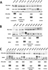
Modification of cysteine-containing mutants of Vph1p by PEG-Mal or NEM. Vacuolar membranes were isolated from the yeast strain MM112 expressing the indicated single cysteine-containing mutants of Vph1p. For the PEG-Mal modification test, 3 μg of vacuolar membrane protein were treated with PEG-Mal in the absence of SDS. For the NEM modification test, 12.5 μg of vacuolar membrane protein were treated sequentially with NEM in the absence of SDS, the NEM was removed, and the samples were then treated with PEG-Mal in the presence of SDS. Samples were separated on SDS-PAGE and Western blotted using the monoclonal antibody 10D7 against Vph1p, as described under “Experimental Procedures.” Modification of Vph1p by PEG-Mal results in a shift in a portion of the Vph1p band by 10–25 kDa. For the NEM modification test, the absence of a shift in mobility of the Vph1p band indicates reactivity toward NEM (see “Results”). A, mutants D710C to A744C; B, mutants G620C to S670C; C, mutants S472C to G602C. Note that the growth phenotype and assembly competence of mutants not shown in Table 2 and Fig. 4 were reported previously (16).
FIGURE 7.
FIGURE 7.
Proposed model of proton-conducting hemichannels in V0. Each helical wheel diagram represents the indicated transmembrane segments viewed from the cytoplasmic side of the membrane. The left panel shows a model of the lumenal hemichannel (shaded blue) with residues contributed from TM3, TM4, and TM7 (also blue). Note that TM7 is oriented so as to optimize the interaction between Ser-740 and His-743 on TM7 (both on the lumenal side of Arg-735) with the lumenally oriented residues on TM3 and TM4. The major disulfide-mediated cross-links that can be formed between TM7 of subunit a and TM3 of subunit c″ (25) are shown by the solid black lines. Following release of a proton from the glutamic acid on the proteolipid subunit into the lumenal hemichannel and stabilization of the negative charge by Arg-735, ATP-driven rotation of the proteolipid ring (shown by the clockwise arrow) brings this glutamic acid into contact with the cytoplasmic hemichannel (shaded yellow in the right panel). Co-incident with this, swiveling of TM7 of subunit a (shown by the counterclockwise arrow) aligns the residues on TM7 that contribute to the cytoplasmic hemichannel (Ser-728, Glu-721, Asn-725, and His-729) with the residues on TM8 that contribute to this hemichannel (Arg-799, Ser-792, His-796, and Glu-789), all shown in yellow. The residues of the lumenal hemichannel are simultaneously misaligned, disrupting the ability of this hemichannel to conduct protons. The cycle is completed by reprotonation of the proteolipid glutamic acid from the cytoplasmic hemichannel.

Similar articles

Cited by

References

    1. Toei M., Saum R., Forgac M. (2010) Biochemistry 49, 4715–4723 - PMC - PubMed
    1. Forgac M. (2007) Nat. Rev. Mol. Cell Biol. 8, 917–929 - PubMed
    1. Kane P. M. (2006) Microbiol. Mol. Biol. Rev. 70, 177–191 - PMC - PubMed
    1. Brown D., Paunescu T. G., Breton S., Marshansky V. (2009) J. Exp. Biol. 212, 1762–1772 - PMC - PubMed
    1. Gruenberg J., van der Goot F. G. (2006) Nat. Rev. Mol. Cell Biol. 7, 495–504 - PubMed

Publication types