

Change in conformation with reduction of alpha-helix content causes loss of neutrophil binding activity in fully cytotoxic Shiga toxin 1
- PMID: 21832076
- PMCID: PMC3186412
- DOI: 10.1074/jbc.M111.255414
Change in conformation with reduction of alpha-helix content causes loss of neutrophil binding activity in fully cytotoxic Shiga toxin 1
Abstract
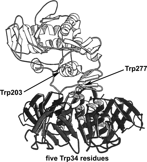
Shiga toxins (Stx) play an important role in the pathogenesis of hemolytic uremic syndrome, a life-threatening renal sequela of human intestinal infection caused by specific Escherichia coli strains. Stx target a restricted subset of human endothelial cells that possess the globotriaosylceramide receptor, like that in renal glomeruli. The toxins, composed of five B chains and a single enzymatic A chain, by removing adenines from ribosomes and DNA, trigger apoptosis and the production of pro-inflammatory cytokines in target cells. Because bacteria are confined to the gut, the toxins move to the kidney through the circulation. Polymorphonuclear leukocytes (PMN) have been indicated as the carriers that "piggyback" shuttle toxins to the kidney. However, there is no consensus on this topic, because not all laboratories have been able to reproduce the Stx/PMN interaction. Here, we demonstrate that conformational changes of Shiga toxin 1, with reduction of α-helix content and exposition to solvent of hydrophobic tryptophan residues, cause a loss of PMN binding activity. The partially unfolded toxin was found to express both enzymatic and globotriaosylceramide binding activities being fully active in intoxicating human endothelial cells; this suggests the presence of a distinct PMN-binding domain. By reviewing functional and structural data, we suggest that A chain moieties close to Trp-203 are recognized by PMN. Our findings could help explain the conflicting results regarding Stx/PMN interactions, especially as the groups reporting positive results obtained Stx by single-step affinity chromatography, which could have preserved the correct folding of Stx with respect to more complicated multi-step purification methods.
Figures

Similar articles
-
Interactions between Shiga toxins and human polymorphonuclear leukocytes.J Leukoc Biol. 2008 Oct;84(4):1019-27. doi: 10.1189/jlb.0308157. Epub 2008 Jul 14. J Leukoc Biol. 2008. PMID: 18625912
-
Shiga toxin 1 and ricin A chain bind to human polymorphonuclear leucocytes through a common receptor.Biochem J. 2010 Nov 15;432(1):173-80. doi: 10.1042/BJ20100455. Biochem J. 2010. PMID: 20809900
-
Lack of specific binding of Shiga-like toxin (verocytotoxin) and non-specific interaction of Shiga-like toxin 2 antibody with human polymorphonuclear leucocytes.Nephrol Dial Transplant. 2007 Mar;22(3):749-55. doi: 10.1093/ndt/gfl688. Epub 2006 Nov 24. Nephrol Dial Transplant. 2007. PMID: 17127697
-
Escherichia coli Shiga toxin.J Nat Toxins. 2000 Aug;9(3):299-313. J Nat Toxins. 2000. PMID: 10994531 Review.
-
Shiga toxins, glycosphingolipid diversity, and endothelial cell injury.Thromb Haemost. 2009 Feb;101(2):252-64. Thromb Haemost. 2009. PMID: 19190807 Review.
Cited by
-
Facing glycosphingolipid-Shiga toxin interaction: dire straits for endothelial cells of the human vasculature.Cell Mol Life Sci. 2013 Feb;70(3):425-57. doi: 10.1007/s00018-012-1060-z. Epub 2012 Jul 6. Cell Mol Life Sci. 2013. PMID: 22766973 Free PMC article. Review.
-
The interactions of human neutrophils with shiga toxins and related plant toxins: danger or safety?Toxins (Basel). 2012 Mar;4(3):157-90. doi: 10.3390/toxins4030157. Epub 2012 Mar 1. Toxins (Basel). 2012. PMID: 22741061 Free PMC article. Review.
-
Association of Shiga toxin glycosphingolipid receptors with membrane microdomains of toxin-sensitive lymphoid and myeloid cells.J Lipid Res. 2013 Mar;54(3):692-710. doi: 10.1194/jlr.M031781. Epub 2012 Dec 17. J Lipid Res. 2013. PMID: 23248329 Free PMC article.
-
Enterohemorrhagic Escherichia coli and a Fresh View on Shiga Toxin-Binding Glycosphingolipids of Primary Human Kidney and Colon Epithelial Cells and Their Toxin Susceptibility.Int J Mol Sci. 2022 Jun 21;23(13):6884. doi: 10.3390/ijms23136884. Int J Mol Sci. 2022. PMID: 35805890 Free PMC article. Review.
-
The structure of the Shiga toxin 2a A-subunit dictates the interactions of the toxin with blood components.Cell Microbiol. 2019 May;21(5):e13000. doi: 10.1111/cmi.13000. Epub 2019 Jan 18. Cell Microbiol. 2019. PMID: 30578712 Free PMC article.
References
-
- Karmali M. A., Petric M., Lim C., Fleming P. C., Arbus G. S., Lior H. (1985) J. Infect. Dis. 151, 775–782 - PubMed
-
- Griffin P. M., Tauxe R. V. (1991) Epidemiol. Rev. 13, 60–98 - PubMed
-
- Caprioli A., Luzzi I., Rosmini F., Pasquini P., Cirrincione R., Gianviti A., Matteucci M. C., Rizzoni G. (1992) J. Infect. Dis. 166, 154–158 - PubMed
Publication types
MeSH terms
Substances
LinkOut - more resources
Full Text Sources
Other Literature Sources
