. 2011 Sep 30;286(39):34244-53.
doi: 10.1074/jbc.M111.241141. Epub 2011 Aug 5.

Polymorphic triple beta-sheet structures contribute to amide hydrogen/deuterium (H/D) exchange protection in the Alzheimer amyloid beta42 peptide

Affiliations

Polymorphic triple beta-sheet structures contribute to amide hydrogen/deuterium (H/D) exchange protection in the Alzheimer amyloid beta42 peptide

Buyong Ma et al. J Biol Chem. .

Abstract

Characterization of the polymorphic structural range of Aβ oligomers is important to the understanding of the mechanisms of toxicity. Yet for highly polymorphic ensembles, experimental structural elucidation is difficult. Here, we use a combination of NMR solvent protection experiments and computational structural screening to identify major species in the amyloid conformational ensemble. We examined the polymorphic pentamer and fibril seeds of Aβ42 and its mutants and compared the theoretical backbone amide protection obtained from simulations with experimental hydrogen/deuterium (H/D) exchange protection ratio. We observed that highly flexible pentamers do not share structural similarities with fibril seed oligomers, except the turn regions. We found that a novel amyloid structural motif of a triple β-sheet, with the N-terminal residues interacting with the core (Lys(17)-Glu(22)) β-sheet region, correlates with H/D exchange protection. The triple β-sheet Aβ42 oligomer has a minimal exposure of hydrophobic residues and is further stabilized by the E22Q (Dutch) mutation in Alzheimer disease. The experimental H/D exchange solvent protection ratio implies that triple β-sheet fibrils and globulomers could coexist in the Aβ42 ensemble, pointing to a broad heterogeneous aggregate population. Our results suggest that an approach that combines computational modeling with NMR protection data can be a useful strategy for obtaining clues to the preferred conformational species of the assemblies in solution and help in alleviating experimental difficulties and consequently possible errors in the exchange data for Aβ42 fibrils.


Figures

FIGURE 1.
FIGURE 1.
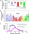
Aβ42 pentamer and fibril have different structure and different H/D exchange protection patterns. A, correlation of the H/D exchange protections for Aβ42 pentamer and fibril is low. B, H/D exchange protections for Aβ42 pentamers can be fitted with the mixture of loop-like structure and several structures with β-hairpins. Because of highly flexible conformations of Aβ42 pentamers, amide hydrogen solvation exposures dominate the H/D exchange process. The solvation exposure values in the y axis are best fit values in Table 1. C, H/D exchange protections for Aβ42 fibril can be fitted with the mixture of triple β-sheet fibril seed oligomer and globulomer. Unlike pentamer, amide hydrogen solvation exposures and backbone hydrogen bonding contribute to H/D exchange protection. The values in x axis correspond to the Kex/Kint in Equation 4 and are obtained with the mixture of GO3 and TF2 models.
FIGURE 2.
FIGURE 2.
Simulations of two disk-like Aβ42 pentamer arrangements indicate that the pentamers are highly flexible. A, disk-like Aβ42 pentamer arrangement based on the suggestion from Ref. . Because of limited interactions among monomers, the arrangement quickly disassociates into monomeric loop structures. B, disk-like Aβ42 pentamer arrangement with β-hairpin structures, which change into loop-like structures with a few β-interactions.
FIGURE 3.
FIGURE 3.
Intramolecular β-hairpin could contribute to the stabilization of Aβ42 pentamers. All these models have the same structure from Val18 to Val36, which fits experimental observation of Phe19–Leu34 interaction in the pentamer. A and E are secondary structures for model m1, which straightforwardly consider that all turns are turns for β-hairpin; B and F are secondary structures for model m2, which consider the region His13–Gln15 and Gly37–Gly38 as bulge in β-strands, making it possible for the C terminus in one monomer to interact with the N terminus in another monomer; C and G are secondary structures for model m3; and D and H are secondary structures for model m4. In the monomer structure of m3 and m4, the C terminus was constructed as an α-helix. The monomers are parallel in the model m3 and anti-parallel in the model m4.
FIGURE 4.
FIGURE 4.
Illustrations of double β-sheet and triple β-sheet fibrils. A, double β-sheet fibril with protofilament contacts in unique Aβ42 interface. B, double β-sheet fibril with protofilaments contacts in Aβ40/42 interface. C, triple β-sheet fibril in Aβ40/42 interface. The triple β-sheet fibril share similar turn regions as in Aβ42 pentamers.
FIGURE 5.
FIGURE 5.
Structures of triple β-sheet models indicate that Aβ peptide can form fibril with minimum exposure of hydrophobic residues. These models are among those simulated in this work. Met35 residues, large balls with sulfur in yellow; Glu22 residues, ball-and-stick with oxygen in red; and Phe4, Tyr10, His14, Val18, Phe20, and Val24 are sticks. A, TF1 model with hydrophobic interactions (HPI) between Phe4 and Phe20; B, TF2 model with HPI between Phe4–Val24 and Tyr10–Val18; C, TF3 model with HPI between Phe4–Val24; D, TF1b model with Met35–Gly37 interaction; E, TF2b model with Met35–Gly37 interaction; and F, TF3E22Q model indicates that buried charges of Glu22 can be neutralized by the E22Q mutation.
FIGURE 6.
FIGURE 6.
Polymorphic ensemble of Aβ can include anti-parallel β-strand arrangements. The models shown here were also simulated in this work. Glu22 and Met35 residues are depicted as ball-and-stick (sulfur, yellow; oxygen, red). A, anti-parallel model AF1 with Val18 aligns with Val18 in neighboring N-terminal strands, and Met35 with Met35 in C-terminal strands; B, anti-parallel model AF2 with the same register as Aβ(16–22) in the D23N mutant Aβ40 fibril (7); in the C-terminal register, Met35 aligns with Gly33 of the neighboring C terminus.
FIGURE 7.
FIGURE 7.
Total number of hydrogen bonds may affect energies and stabilities of oligomers. A, total effective energies for three group of oligomers. The models with blue bar are double β-sheet, red bars are triple β-sheet, and the green bars are anti-parallel models. B, total number of hydrogen bonds for three type of oligomers as follows: double β-sheet F1d (blue), triple β-sheet TF1b (red), and antiparallel AF2 models (green). C, E22Q mutation decreases the hydrogen bonds for double β-sheet structure but increases the hydrogen bonds for triple β-sheet structure.
FIGURE 8.
FIGURE 8.
Balances among electrostatic interaction and hydrophobic interaction affect stabilities of fibrils for wild type and E22Q mutants. A, antiparallel model AF2 has the most favorable electrostatic interactions. However, E22Q mutation make the triple β-sheet model TF2b-E22Q also have close electrostatic interactions as the AF2-E22Q model. The electrostatic interactions in the double β-sheet model F1d-E22Q are not as favorable as other two models. B, triple β-sheet fibril model TF2 decreases the exposure of hydrophobic residues, and the E22Q mutation leads to a greater burial of hydrophobic residues. The models with blue bar are double β-sheet, red bars are triple β-sheet, yellow bars are cited in Ref. , and the green bars are anti-parallel models. C, E22Q has a shorter distance between the N-terminal and central β-sheet regions. The unit of distance is Å.

Similar articles

Cited by

References

    1. Lührs T., Ritter C., Adrian M., Riek-Loher D., Bohrmann B., Döbeli H., Schubert D., Riek R. (2005) Proc. Natl. Acad. Sci. U.S.A. 102, 17342–17347 - PMC - PubMed
    1. Frydman-Marom A., Rechter M., Shefler I., Bram Y., Shalev D. E., Gazit E. (2009) Angew. Chem. Int. Ed. Engl. 48, 1981–1986 - PubMed
    1. Yan L. M., Velkova A., Tatarek-Nossol M., Andreetto E., Kapurniotu A. (2007) Angew. Chem. Int. Ed. Engl. 46, 1246–1252 - PubMed
    1. Schmidt M., Sachse C., Richter W., Xu C., Fändrich M., Grigorieff N. (2009) Proc. Natl. Acad. Sci. U.S.A. 106, 19813–19818 - PMC - PubMed
    1. Olofsson A., Lindhagen-Persson M., Sauer-Eriksson A. E., Ohman A. (2007) Biochem. J. 404, 63–70 - PMC - PubMed

Publication types