

Expression of ezrin in human embryonic, fetal, and normal adult tissues
- PMID: 21832146
- PMCID: PMC3261628
- DOI: 10.1369/0022155411418661
Expression of ezrin in human embryonic, fetal, and normal adult tissues
Abstract
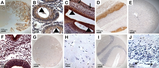
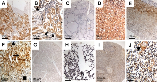
Ezrin, which cross-links the cytoskeleton and plasma membrane, was involved in a wide variety of cellular processes. Here, to investigate the distribution of ezrin, tissue microarray technology was employed to perform immunohistochemical experiments on human embryos, fetuses at 4 to 22 weeks' gestation, and adult tissue specimens. Results showed that ezrin was widely expressed in the gastrointestinal tract throughout the human developmental stages studied. At 6 to 8 weeks' gestation, ezrin was found in epithelial cells, and this staining pattern was particularly pronounced in the brush border of mature absorptive cells lining the villus in later developmental stages and adult tissues. Throughout neural development, ezrin was only expressed in the neural tube at 4 weeks' gestation. Ezrin was also detected in the cortex and medulla of the adrenal gland at 8 to 12 weeks' gestation, whereas its immunoreactivity was increased from the zona glomerulosa through the zona reticularis and was essentially undetectable in the adrenal medulla of adult tissues. Significant expression of ezrin was seen throughout development in the kidney, spleen, lymph nodes, and cells of stratified squamous epithelia. However, ezrin was undetectable in lung, liver, heart, and blood vessels. These results demonstrated that the expression pattern of ezrin was highly time specific and tissue specific.
Conflict of interest statement
The author(s) declared no potential conflicts of interest with respect to the authorship and/or publication of this article.
Figures
Similar articles
-
Fascin expression in human embryonic, fetal, and normal adult tissue.J Histochem Cytochem. 2008 Feb;56(2):193-9. doi: 10.1369/jhc.7A7353.2007. Epub 2007 Nov 12. J Histochem Cytochem. 2008. PMID: 17998567 Free PMC article.
-
Expression of NGAL and NGALR in human embryonic, fetal and normal adult tissues.Mol Med Rep. 2012 Oct;6(4):716-22. doi: 10.3892/mmr.2012.980. Epub 2012 Jul 6. Mol Med Rep. 2012. PMID: 22797813
-
Differential expression of HSPA1 and HSPA2 proteins in human tissues; tissue microarray-based immunohistochemical study.Histochem Cell Biol. 2011 Apr;135(4):337-50. doi: 10.1007/s00418-011-0791-5. Epub 2011 Mar 4. Histochem Cell Biol. 2011. PMID: 21373891 Free PMC article.
-
Immunohistochemical profile of ezrin and radixin in human liver epithelia during fetal development and pediatric cholestatic diseases.Clin Res Hepatol Gastroenterol. 2013 Apr;37(2):142-51. doi: 10.1016/j.clinre.2013.02.001. Epub 2013 Mar 16. Clin Res Hepatol Gastroenterol. 2013. PMID: 23507543
-
Does fetal antigen 1 (FA1) identify cells with regenerative, endocrine and neuroendocrine potentials? A study of FA1 in embryonic, fetal, and placental tissue and in maternal circulation.Differentiation. 2000 Aug;66(1):49-59. doi: 10.1046/j.1432-0436.2000.066001049.x. Differentiation. 2000. PMID: 10997592
Cited by
-
Transcriptomics analysis identified ezrin as a potential druggable target in cervical and gastric cancer cells.Clinics (Sao Paulo). 2024 Jul 6;79:100422. doi: 10.1016/j.clinsp.2024.100422. eCollection 2024. Clinics (Sao Paulo). 2024. PMID: 38972247 Free PMC article.
-
Expression of ezrin and moesin in primary breast carcinoma and matched lymph node metastases.Clin Exp Metastasis. 2017 Jun;34(5):333-344. doi: 10.1007/s10585-017-9853-y. Epub 2017 Jun 17. Clin Exp Metastasis. 2017. PMID: 28624994
-
Comparison of the broncoalveolar lavage fluid proteomics between foals and adult horses.PLoS One. 2023 Sep 5;18(9):e0290778. doi: 10.1371/journal.pone.0290778. eCollection 2023. PLoS One. 2023. PMID: 37669266 Free PMC article.
-
Post-translational modifications of ezrin: a crucial regulator for diseases.Amino Acids. 2025 Aug 9;57(1):40. doi: 10.1007/s00726-025-03472-3. Amino Acids. 2025. PMID: 40782214 Free PMC article. Review.
-
Two Sides of the Coin: Ezrin/Radixin/Moesin and Merlin Control Membrane Structure and Contact Inhibition.Int J Mol Sci. 2019 Apr 23;20(8):1996. doi: 10.3390/ijms20081996. Int J Mol Sci. 2019. PMID: 31018575 Free PMC article. Review.
References
-
- Barilá D, Murgia C, Nobili F, Perozzi G. 1995. Transcriptional regulation of the ezrin gene during rat intestinal development and epithelial differentiation. Biochim Biophys Acta. 1263:133–140 - PubMed
-
- Berryman M, Franck Z, Bretscher A. 1993. Ezrin is concentrated in the apical microvilli of a wide variety of epithelial cells whereas moesin is found primarily in endothelial cells. J Cell Sci. 105:1025–1043 - PubMed
Publication types
MeSH terms
Substances
LinkOut - more resources
Full Text Sources
Other Literature Sources
