

Chronic UVR causes increased immunostaining of CD44 and accumulation of hyaluronan in mouse epidermis
- PMID: 21832148
- PMCID: PMC3201128
- DOI: 10.1369/0022155411417874
Chronic UVR causes increased immunostaining of CD44 and accumulation of hyaluronan in mouse epidermis
Abstract
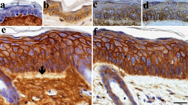
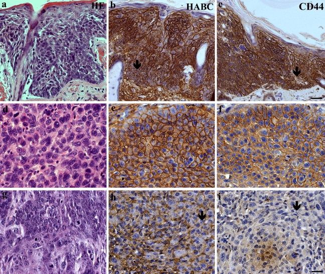
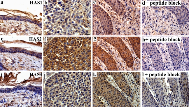
Chronic intense UV radiation is the main cause of epidermal tumors. Because hyaluronan (HA), a large extracellular polysaccharide, is known to promote malignant growth, hyaluronan expression was studied in a model in which long-term UV radiation (UVR) induces epidermal tumors. Mouse back skin was exposed three times a week for 10.5 months to UVR corresponding to one minimal erythema dose, processed for histology, and stained for hyaluronan and the hyaluronan receptor CD44. This exposure protocol caused epidermal hyperplasia in most of the animals; tumors, mainly squamous cell carcinomas (SCCs), were found in ~20% of the animals. Specimens exposed to UVR showed increased hyaluronan and CD44 staining throughout the epidermal tissue. In hyperplastic areas, hyaluronan and CD44 stainings correlated positively with the degree of hyperplasia. Well-differentiated SCCs showed increased hyaluronan and CD44 staining intensities, whereas poorly differentiated tumors and dysplastic epidermis showed areas where HA and CD44 were locally reduced. The findings indicate that HA and CD44 increase in epidermal keratinocytes in the premalignant hyperplasia induced by UV irradiation and stay elevated in dysplasia and SCC, suggesting that the accumulation of hyaluronan and CD44 is an early marker for malignant transformation and may be a prerequisite for tumor formation.
Conflict of interest statement
The author(s) declared no potential conflicts of interest with respect to the authorship and publication of this article.
Figures
Similar articles
-
Osteopontin facilitates ultraviolet B-induced squamous cell carcinoma development.J Dermatol Sci. 2014 Aug;75(2):121-32. doi: 10.1016/j.jdermsci.2014.05.002. Epub 2014 May 21. J Dermatol Sci. 2014. PMID: 24888687 Free PMC article.
-
Hyaluronan, CD44 and versican in epidermal keratinocyte tumours.Br J Dermatol. 2003 Jan;148(1):86-94. doi: 10.1046/j.1365-2133.2003.05028.x. Br J Dermatol. 2003. PMID: 12534600
-
Low dose ultraviolet B irradiation increases hyaluronan synthesis in epidermal keratinocytes via sequential induction of hyaluronan synthases Has1-3 mediated by p38 and Ca2+/calmodulin-dependent protein kinase II (CaMKII) signaling.J Biol Chem. 2013 Jun 21;288(25):17999-8012. doi: 10.1074/jbc.M113.472530. Epub 2013 May 3. J Biol Chem. 2013. PMID: 23645665 Free PMC article.
-
Selective Hyaluronan-CD44 Signaling Promotes miRNA-21 Expression and Interacts with Vitamin D Function during Cutaneous Squamous Cell Carcinomas Progression Following UV Irradiation.Front Immunol. 2015 May 13;6:224. doi: 10.3389/fimmu.2015.00224. eCollection 2015. Front Immunol. 2015. PMID: 26029210 Free PMC article. Review.
-
UV-Induced Molecular Signaling Differences in Melanoma and Non-melanoma Skin Cancer.Adv Exp Med Biol. 2017;996:27-40. doi: 10.1007/978-3-319-56017-5_3. Adv Exp Med Biol. 2017. PMID: 29124688 Review.
Cited by
-
HAS3-induced accumulation of hyaluronan in 3D MDCK cultures results in mitotic spindle misorientation and disturbed organization of epithelium.Histochem Cell Biol. 2012 Feb;137(2):153-64. doi: 10.1007/s00418-011-0896-x. Epub 2011 Dec 8. Histochem Cell Biol. 2012. PMID: 22159845
-
Pterygium-The Good, the Bad, and the Ugly.Cells. 2021 Jun 22;10(7):1567. doi: 10.3390/cells10071567. Cells. 2021. PMID: 34206333 Free PMC article. Review.
-
Hyaluronan accumulation is associated with reduced hyaluronidase expression in renal cell carcinoma, with CD44, HAS1, and HYAL2 emerging as prognostic markers.J Pathol Clin Res. 2025 Jul;11(4):e70035. doi: 10.1002/2056-4538.70035. J Pathol Clin Res. 2025. PMID: 40528762 Free PMC article.
-
Osteopontin facilitates ultraviolet B-induced squamous cell carcinoma development.J Dermatol Sci. 2014 Aug;75(2):121-32. doi: 10.1016/j.jdermsci.2014.05.002. Epub 2014 May 21. J Dermatol Sci. 2014. PMID: 24888687 Free PMC article.
-
CD44 Immunoexpression in the Progression of Actinic Keratosis and Cutaneous Squamouscarcinoma.Curr Health Sci J. 2017 Jul-Sep;43(3):241-245. doi: 10.12865/CHSJ.43.03.10. Epub 2017 Sep 28. Curr Health Sci J. 2017. PMID: 30595883 Free PMC article.
References
-
- Armstrong BK, Kricker A. 2001. The epidemiology of UV induced skin cancer. J Photochem Photobiol B. 63:8–18 - PubMed
-
- Averbeck M, Gebhardt CA, Voigt S, Beilharz S, Anderegg U, Termeer CC, Sleeman JP, Simon JC. 2007. Differential regulation of hyaluronan metabolism in the epidermal and dermal compartments of human skin by UVB irradiation. J Invest Dermatol. 127:687–697 - PubMed
-
- Bakkers J, Kramer C, Pothof J, Quaedvlieg NE, Spaink HP, Hammerschmidt M. 2004. Has2 is required upstream of Rac1 to govern dorsal migration of lateral cells during zebrafish gastrulation. Development. 131:525–537 - PubMed
-
- Bourguignon LY, Gilad E, Peyrollier K. 2007. Heregulin-mediated ErbB2-ERK signaling activates hyaluronan synthases leading to CD44-dependent ovarian tumor cell growth and migration. J Biol Chem. 282:19426–19441 - PubMed
Publication types
MeSH terms
Substances
LinkOut - more resources
Full Text Sources
Other Literature Sources
Medical
Molecular Biology Databases
Research Materials
Miscellaneous
