. 2011 Oct;22(19):3584-94.
doi: 10.1091/mbc.E11-01-0012. Epub 2011 Aug 10.

Phosphorylation of the Ndc80 complex protein, HEC1, by Nek2 kinase modulates chromosome alignment and signaling of the spindle assembly checkpoint

Affiliations

Phosphorylation of the Ndc80 complex protein, HEC1, by Nek2 kinase modulates chromosome alignment and signaling of the spindle assembly checkpoint

Randy Wei et al. Mol Biol Cell. 2011 Oct.

Abstract

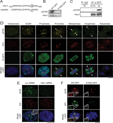
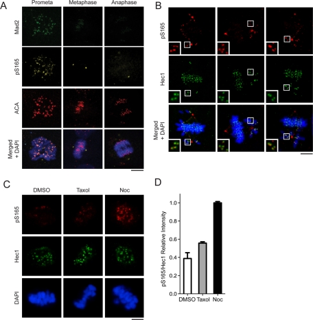
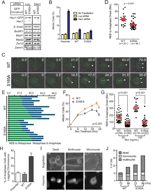
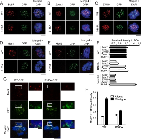
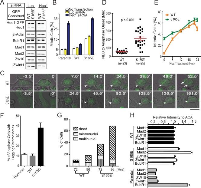
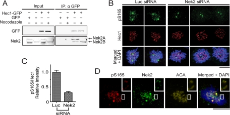
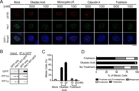
The spindle assemble checkpoint (SAC) is critical for accurate chromosome segregation. Hec1 contributes to chromosome segregation in part by mediating SAC signaling and chromosome alignment. However, the molecular mechanism by which Hec1 modulates checkpoint signaling and alignment remains poorly understood. We found that Hec1 serine 165 (S165) is preferentially phosphorylated at kinetochores. Phosphorylated Hec1 serine 165 (pS165) specifically localized to kinetochores of misaligned chromosomes, showing a spatiotemporal distribution characteristic of SAC molecules. Expressing an RNA interference (RNAi)-resistant S165A mutant in Hec1-depleted cells permitted normal progression to metaphase, but accelerated the metaphase-to-anaphase transition. The S165A cells were defective in Mad1 and Mad2 localization to kinetochores, regardless of attachment status. These cells often entered anaphase with lagging chromosomes and elicited increased segregation errors and cell death. In contrast, expressing S165E mutant in Hec1-depleted cells triggered defective chromosome alignment and severe mitotic arrest associated with increased Mad1/Mad2 signals at prometaphase kinetochores. A small portion of S165E cells eventually bypassed the SAC but showed severe segregation errors. Nek2 is the primary kinase responsible for kinetochore pS165, while PP1 phosphatase may dephosphorylate pS165 during SAC silencing. Taken together, these results suggest that modifications of Hec1 S165 serve as an important mechanism in modulating SAC signaling and chromosome alignment.


Figures

FIGURE 1:
FIGURE 1:
Generation of phospho-specific antibody recognizing Hec1 S165 phosphorylation during mitosis. (A) Diagram of Hec1 S165 phosphopeptide antigen used for generating phospho-specific antibody. (B) Western blot analysis of lysates from Hec1-WT-GFP– or Hec1-S165A-GFP–expressing cells using pS165 antibody. The same blot was stripped and reprobed with Hec1 (9G3) monoclonal antibody. (C) U2OS cells enriched at G1/S (double-thymidine block) or M phase (nocodazole), for which anti-pS165 antibody was used to blot immunoprecipitated Hec1-WT-GFP. The same blot was stripped and reprobed with anti-Hec1. (D) Unsynchronized MCF10A cells were stained with anti-pS165 (yellow), anti-centromere antibody (ACA; red), anti-Histone3 pSer10 antibodies (green), and DAPI (blue). Arrows indicate spindle poles. (E) Immunofluorescence staining with anti-pS165 (green) and ACAs (red) of U2OS cells transfected with Hec1 or luciferase siRNA. Inset image shows one pair of kinetochores. (F) pS165 staining is revealed in U2OS cells rescued with RNAi-resistant Hec1-WT-GFP, but not S165A mutant, when endogenous Hec1 is depleted. Insets show a pair of kinetochores. Scale bars: 5 μm.
FIGURE 2:
FIGURE 2:
Phosphorylation of Hec1 S165 occurs specifically on kinetochores of misaligned chromosomes. (A) pS165 costaining with Mad2 in mitotic U2OS cells (prometaphase, metaphase, and anaphase). (B) pS165 costaining with Hec1 in mitotic MCF10A cells with misaligned chromosomes. Insets show magnified images of boxed regions. (C) pS165 costaining with Hec1 in U2OS cells arrested for 2 h with 10 μM MG132 prior to treatment with dimethyl sulfoxide (DMSO), Taxol (final concentration, 10 μM), or nocodazole (300 nM) for 1 h. (D) Relative fluorescence intensity of pS165 vs. total Hec1 in each cell population from (C) images (mean ± SEM; n > 200 kinetochores from seven individual cells). All images were captured under the same exposure settings. For all images, chromosomes were stained with DAPI (blue). Scale bars: 5 μm.
FIGURE 3:
FIGURE 3:
Expression of Hec1 S165A results in shortened mitotic duration and increased chromosomal segregation errors. (A) Western blot analysis of Hec1-depleted cells expressing RNAi-resistant Hec1-WT-GFP or Hec1-S165A-GFP. Luciferase siRNA was used as a control. The expressions of BubR1, Mad1, Mad2, Zw10, and Zwint1 were also examined. β-Actin served as an internal loading control. (B) Mitotic index of U2OS parental cells and U2OS cells ectopically expressing RNAi-resistant Hec1-WT-GFP or Hec1-S165A-GFP. Cells were either transfected with luciferase or Hec1 siRNA, or not transfected at all. About 1000 cells were collected for each sample from two separate experiments. (C) Time-lapse microscopy of WT and S165A cell during mitosis. Images were captured at 3.5-min intervals. Triangles indicate chromosomes aligned along the metaphase plate. Arrows indicate separated sister chromatids. Scale bar: 10 μm. (D) Quantification of time from NEB to anaphase onset in WT and S165A cells without nocodazole. (E) Duration of time that WT or S165A cells were undergoing NEB-to-metaphase onset and metaphase-to-anaphase onset. (F) Percentage of mitotic cells expressing WT or S165A mutant after depletion of the endogenous Hec1 and treatment with 50 ng/ml nocodazole. Cells were randomly photographed at the designated time point using fluorescence and phase-contrast microscopy. About 500 cells were collected for each sample and time point. (G) Quantification of time from NEB-to-anaphase onset in WT and S165A cells with 20 or 100 ng/ml of nocodazole. (mean ± SEM; images of cells collected from two independent experiments; p < 0.001). (H) Percentage of WT or S165A cells with lagging chromosomes during anaphase 48 h after Hec1 siRNA transfection. (I) Representative images of interphase nuclei stained with Hoechst 33342 (top) and their corresponding bright field images (bottom). Scale bar: 10 μm. (J) Quantification of multinucleated, micronucleated, and dead cells 72 and 96 h after depletion of endogenous Hec1 with siRNA (mean ± SEM; n > 400 cells per sample).
FIGURE 4:
FIGURE 4:
Expression of Hec1 S165A impairs recruitment of Mad1 and Mad2 to kinetochores. (A–E) WT and S165A cells were immunostained with antibodies against BubR1 (A), Zwint1 (B), ZW10 (C), Mad1 (D), and Mad2 (E) after depletion of endogenous Hec1. Scale bar: 5 μm. (F) Quantification of SAC-staining intensities at kinetochores (normalized for ACA staining; mean ± SEM; n > 150 kinetochores from 10 different cells). (G) Mad2 staining in U2OS cells rescued with Hec1-WT-GFP or the S165A mutant when endogenous Hec1 is depleted. Insets show magnified images of boxed regions. (H) Quantification of Mad2 fluorescence intensity on aligned and misaligned chromosomes. For all images, chromosomes were stained with DAPI (blue). Scale bars: 5 μm.
FIGURE 5:
FIGURE 5:
Expression of Hec1 S165E prolongs mitotic duration, increases segregation errors, and causes cell death. (A) Western blot analysis of Hec1-depleted cells expressing RNAi-resistant Hec1-WT-GFP or Hec1-S165E-GFP. Luciferase siRNA was used as a control. The expressions of BubR1, Mad1, Mad2, Zw10, and Zwint1 were also examined with their specific antibodies. β-actin served as a loading control. (B) Mitotic index of U2OS parental cells and U2OS cells ectopically expressing RNAi-resistant Hec1-WT-GFP or Hec1-S165E-GFP. Cells were either transfected with luciferase or Hec1 siRNA, or untransfected. About 1000 cells were collected for each sample from two separate experiments. (C) Time-lapse microscopy of WT and S165E cell during mitosis. Images were captured at 3.5-min intervals. Triangles indicate chromosomes aligned along the metaphase plate. Arrows indicate separated sister chromatids. Scale bar: 10 μm. (D) Quantification of time from NEB-to-anaphase onset of WT and S165E cells without nocodazole. (mean ± SEM; images collected from two independent experiments; p < 0.001). (E) Percentage of mitotic cells expressing WT or S165E mutant after depletion of the endogenous Hec1 with siRNA and subsequent treatment with 50 ng/ml nocodazole. Cells were randomly photographed at the designated time point using fluorescence and phase-contrast microscopy. About 500 cells were collected for each sample and time point. (F) Percentage of WT or S165A cells with lagging chromosomes during anaphase 48 h after Hec1 siRNA transfection. (G) Quantification of multinucleated, micronucleated, and dead cells 72 and 96 h after depletion of endogenous Hec1 (mean ± SEM; n > 400 cells per sample). (H) Quantification of SAC-staining intensities at kinetochores (normalized for ACA staining; mean ± SEM; n > 150 kinetochores from 10 different cells).
FIGURE 6:
FIGURE 6:
Nek2 phosphorylates Hec1 on S165. (A) In untreated GFP and Hec1-WT–expressing U2OS cells, Nek2A and Nek2B are expressed at comparable levels, and coimmunoprecipitation of Hec1-WT pulled down both Nek2A and Nek2B. On nocodazole treatment, Nek2A but not Nek2B levels were reduced, and only Nek2B coimmunoprecipitated with Hec1-WT. (B) pS165 staining is present in U2OS cells transfected with luciferase siRNA, but not when transfected with Nek2 siRNA. Cells were stained with anti-pS165 (red), anti-Hec1 antibodies (green), and DAPI (blue). (C) Quantification of pS165-staining intensities at kinetochores (normalized for total Hec1) in luciferase or Nek2 siRNA-treated U2OS cells (mean ± SEM; n > 200 kinetochores from seven individual cells.) All images were captured under the same exposure settings. (D) Nek2 and pS165 colocalized at kinetochores of U2OS cells. Cells were costained with anti-pS165 (red) and anti-Nek2 antibodies (green), anti-ACA (yellow), and DAPI (blue). Inset is higher magnification image of boxed region. Scale bars: 5 μm.
FIGURE 7:
FIGURE 7:
Inhibition of PP1 phosphatase preserves Hec1 pS165 signal. (A) MCF10A cells were immunostained with anti-pS165 antibodies (red) and anti-Hec1 (9G3), antibodies (green), using prefixation extraction buffer containing either 500 nM or 100 nM of specific phosphatase inhibitor or mock inhibitor (DMSO). Cells were counterstained with DAPI (blue). Scale bar: 5 μm. (B) Ectopically expressed Hec1-WT-GFP was coimmunoprecipitated with endogenous PP1α and PP1γ by anti-GFP antibody. (C) Mitotic index of U2OS cells treated for 18 h with 10 or 20 nM of either okadaic acid or fostriecin. About 1000 cells were collected for each sample from two separate experiments. (D) Mitotic distribution of different stages of U2OS cells following treatment with 20 nM okadaic acid or 20 nM fostriecin for 18 h.

Similar articles

Cited by

References

    1. Alushin GM, Ramey VH, Pasqualato S, Ball DA, Grigorieff N, Musacchio A, Nogales E. The Ndc80 kinetochore complex forms oligomeric arrays along microtubules. Nature. 2010;467:805–810. - PMC - PubMed
    1. Brito DA, Rieder CL. The ability to survive mitosis in the presence of microtubule poisons differs significantly between human nontransformed (RPE-1) and cancer (U2OS, HeLa) cells. Cell Motil Cytoskeleton. 2009;66:437–447. - PMC - PubMed
    1. Buffin E, Lefebvre C, Huang J, Gagou ME, Karess RE. Recruitment of Mad2 to the kinetochore requires the Rod/Zw10 complex. Curr Biol. 2005;15:856–861. - PubMed
    1. Burds AA, Lutum AS, Sorger PK. Generating chromosome instability through the simultaneous deletion of Mad2 and p53. Proc Natl Acad Sci USA. 2005;102:11296–11301. - PMC - PubMed
    1. Burns JC, Friedmann T, Driever W, Burrascano M, Yee JK. Vesicular stomatitis virus G glycoprotein pseudotyped retroviral vectors: concentration to very high titer and efficient gene transfer into mammalian and nonmammalian cells. Proc Natl Acad Sci USA. 1993;90:8033–8037. - PMC - PubMed

Publication types

MeSH terms

LinkOut - more resources