

Mild sensory stimulation reestablishes cortical function during the acute phase of ischemia
- PMID: 21832179
- PMCID: PMC3162364
- DOI: 10.1523/JNEUROSCI.1741-11.2011
Mild sensory stimulation reestablishes cortical function during the acute phase of ischemia
Abstract
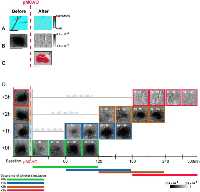
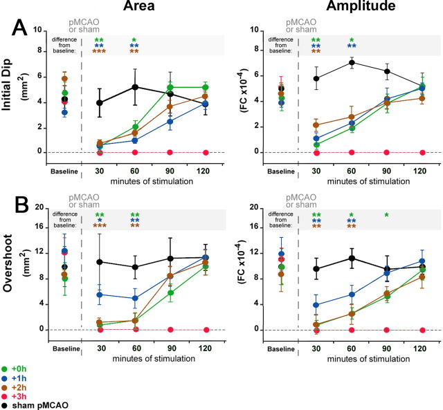
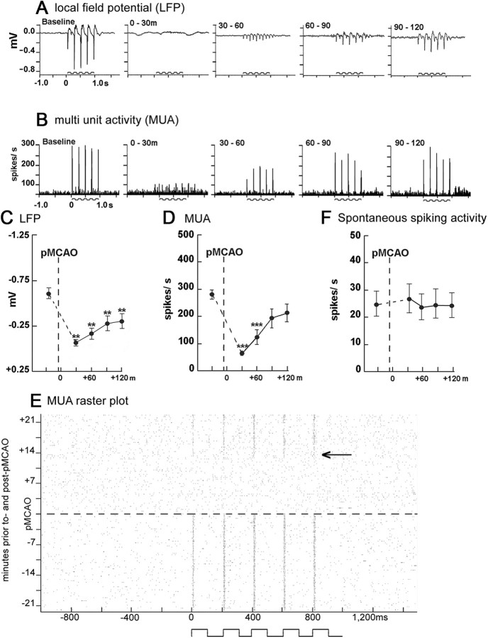
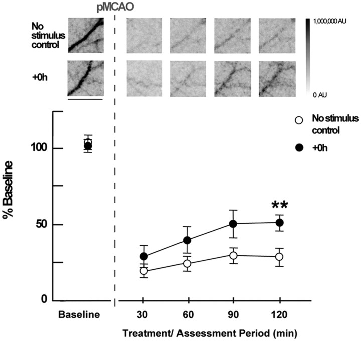
When delivered within 1 and in most cases 2 h of permanent middle cerebral artery occlusion (pMCAO), mild sensory stimulation (intermittent single whisker stimulation) was shown to be completely neuroprotective 24 h after pMCAO in a rodent model of ischemic stroke, according to assessment with multiple techniques (Lay et al., 2010). The acute effect of stimulation treatment on the ischemic cortex, however, has yet to be reported. Here we characterize cortical function and perfusion during the 120 min whisker stimulation period in four experimental groups with treatment initiated 0, 1, 2 (protected groups), or 3 h (unprotected group) post-pMCAO using multiple techniques. According to functional imaging, a gradual return of evoked whisker functional representation to baseline levels was initiated with treatment onset and completed within the treatment period. Evoked neuronal activity and reperfusion to the ischemic area also showed a gradual recovery in protected animals. Surprisingly, a similar recovery profile was observed in response to treatment in all protected animals, regardless of treatment onset time. Nonstimulated pMCAO control group data demonstrate that reperfusion is not spontaneous. This makes the complete protection observed in the majority of animals stimulated at 2 h post-pMCAO even more surprising, as these animals recovered despite having been in a severely ischemic state for two full hours. In summary, when delivered within a 2 h window post-pMCAO, whisker stimulation treatment initiated reperfusion and a gradual recovery of cortical function that was completed or nearly completed within the treatment period.
Figures
References
-
- Bederson JB, Pitts LH, Tsuji M, Nishimura MC, Davis RL, Bartkowski H. Rat middle cerebral artery occlusion: evaluation of the model and development of a neurologic examination. Stroke. 1986;17:472–476. - PubMed
-
- Briers JD, Webster S. Quasi real-time digital version of single-exposure photography for full-field monitoring of velocity or flow-fields. Opt Commun. 1995:36–42.
-
- Brint S, Jacewicz M, Kiessling M, Tanabe J, Pulsinelli W. Focal brain ischemia in the rat: methods for reproducible neocortical infarction using tandem occlusion of the distal middle cerebral and ipsilateral common carotid arteries. J Cereb Blood Flow Metab. 1988;8:474–485. - PubMed
Publication types
MeSH terms
Grants and funding
LinkOut - more resources
Full Text Sources