. 2011 Oct 13;118(15):4274-84.
doi: 10.1182/blood-2011-03-339531. Epub 2011 Aug 10.

Tetraspanin CD151 maintains vascular stability by balancing the forces of cell adhesion and cytoskeletal tension

Affiliations

Tetraspanin CD151 maintains vascular stability by balancing the forces of cell adhesion and cytoskeletal tension

Feng Zhang et al. Blood. .

Erratum in

  • Blood. 2014 Jun 12;123(24):3843

Abstract

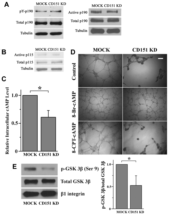
Tetraspanin CD151 is highly expressed in endothelial cells and regulates pathologic angiogenesis. However, the mechanism by which CD151 promotes vascular morphogenesis and whether CD151 engages other vascular functions are unclear. Here we report that CD151 is required for maintaining endothelial capillary-like structures formed in vitro and the integrity of endothelial cell-cell and cell-matrix contacts in vivo. In addition, vascular permeability is markedly enhanced in the absence of CD151. As a global regulator of endothelial cell-cell and cell-matrix adhesions, CD151 is needed for the optimal functions of various cell adhesion proteins. The loss of CD151 elevates actin cytoskeletal traction by up-regulating RhoA signaling and diminishes actin cortical meshwork by down-regulating Rac1 activity. The inhibition of RhoA or activation of cAMP signaling stabilizes CD151-silenced or -null endothelial structure in vascular morphogenesis. Together, our data demonstrate that CD151 maintains vascular stability by promoting endothelial cell adhesions, especially cell-cell adhesion, and confining cytoskeletal tension.


Figures

Figure 1
Figure 1
CD151 reinforces vascular stability and regulates vascular permeability. (A) Loss of CD151 expression disrupted EC capillary-like structures on Matrigel. HMEC-MOCK and -CD151 KD (top) or MLEC-WT and -CD151 KO (bottom) cells were plated on Matrigel and photographed with an Olympus CK2 inverted microscope under a 4×/0.10 NA objective equipped with a microscope digital camera (DCM500) at the indicated time points. The cable-enclosed regions were counted and compared. Bar represents 250 μm. *P < .01. (B) CD151 ablation results in increased vascular permeability in mice. Evans blue dye (30 mg/kg in PBS) was injected intravenously through the retro-orbital sinus into 12-week-old male mice. Mustard oil in mineral oil (5% volume/volume) or mineral oil alone was applied to the dorsal and ventral surfaces of the ears twice, at 0 minutes and 15 minutes later. After 30 minutes of circulation, the dye leakage area at the ventral surface of the ear was imaged with a Nikon SMZ1500 dissecting microscope equipped with a Nikon DXM1200 digital camera and the content of dye in ears was determined after the extraction with formamide overnight at 55°C. Shown are representative images (top) and quantitative results from WT and CD151 KO mice from the Miles assay. n = 15. Bar represents 1 mm. **P < .05.
Figure 2
Figure 2
CD151 up-regulates endothelial cell-matrix adhesiveness. (A) Cell-matrix adhesion assay. HMEC transductants were seeded on ECM substrate-coated wells in triplicates and allowed to settle at 37°C for 35 minutes. Nonadherent cells were then removed by gentle washing. The adhered cells were counted. *P < .01. (B) Traction force microscopy. Cells were plated on FN-, LN 111-, or LN 332-conjugated fluorescent bead-embedded polyacrylamide gels. Traction forces exerted by the cells were measured as described in “Traction force microscopy.” Left: Phase-contrast images and traction field of cells on FN-conjugated polyarylamide gels. Bar represents 10 μm. Right: Maximum and average perimeter traction forces of the HMEC transductants on FN, LN 111, or LN 332. The magnitudes of maximum and average perimeter traction forces were compared with a nonparametric Mann-Whitney test. **P < .05. (C) CD151 silencing attenuates the formation and maturation of focal adhesions. HMEC transductants, after 2-day culture, were fixed, permeabilized, and incubated with vinculin mAb, followed by AlexaFluor-488–conjugated secondary Ab and phalloidin–AlexaFluor-594 staining. Staining was imaged with a Zeiss LSM510 confocal fluorescence microscope under a 100×/1.4 NA oil objective. Bar represents 10 μm. (D) TIRF microscopy. HMEC transductants were fixed without permeabilization, probed with α3 integrin mAb and AlexaFluro-488–conjugated secondary Ab, and visualized by TIRF microscopy. Bar represents 15 μm. The size and numbers of fluorescent particles of α3 integrin and CD9 from the individual transductants (n = 20) were analyzed and compared. (E) Silencing of CD151 expression elevates detergent solubility of β1 integrin. HMEC transductants were lysed with 0.05% Triton X-100 in HEPES buffer. After ultracentrifugation, supernatants were used as soluble fractions. Pellets were solubilized in 1× Laemmli sample buffer and used as the insoluble fractions. Integrin β1 in both fractions as well as whole cell lysates were subjected to SDS-PAGE and then detected by immunoblotting using TS2/16 mAb. Tubulin from whole cell lysates was used as loading control. (F) TS2/16 partially rescues the defects in maintenance of capillary structures in CD151-silenced ECs. HMEC transductants were incubated with 1 μg/mL β1 integrin-activating mAb TS2/16 on ice for 1 hour and were plated on Matrigel in the presence of TS2/16. The capillary-like structures were imaged and quantified. An isotype-matched antihuman CD71 mAb served as the negative control. Bar represents 250 μm. (G) Active β1 integrins were unaltered on CD151 silencing. The levels of activated β1 integrins on HMECs were measured by flow cytometry using mAb AG89. (H) The dissociation of ECs from the basement membrane (BM) and splitting of BM in the absence of CD151. Twelve-week-old male CD151 KO (n = 4) and littermate WT (n = 4) mice were perfused and fixed with 2.5% glutaraldehyde. The aortas were isolated, sectioned transversely, and processed for transmission electron microscopy. The red asterisks indicate the space where ECs were detached from BM. Bar represents 250 nm.
Figure 3
Figure 3
CD151 is needed for proper endothelial cell-cell adhesion. (A) Cell aggregation assay. A total of 2 × 10⁴ cells were seeded into 30-μL hanging drop cultures in either complete or Ca2+-depleted media and allowed to aggregate at 37°C overnight. After passing the cell cluster 10 times through a 200-μL pipette tip, cell clusters were imaged, and the degree of dissociation of the aggregates was quantified using ImageJ 1.42q (published by Wayne Rasband, National Institutes of Health) software analysis. Representative images of aggregation in untreated cells (left panel) and the quantitative results (right panel). Bar represents 100 μm. *P < .01. (B) AJ complexes are mislocalized in CD151-silenced ECs. HUVEC transductants were cultured for 4 days to confluence. After 6-hour starvation, HUVEC monolayers were fixed, permeabilized, and stained with specific Abs. The distributions of VE-cadherin and β-catenin were visualized using a Zeiss LSM510 confocal fluorescence microscope under a 100×/1.4 NA oil objective. Bar represents 10 μm. (C) Deficient formation and maturation of adhesion zipper on CD151 silencing. HMEC transductants were fixed without permeabilization, probed with CD9 mAb and AlexaFluro-488–conjugated secondary Ab, and visualized by TIRF microscopy. Bar represents 15 μm. (D) Abnormal endothelial cell-cell adhesion in lung vessels of CD151 KO mice. Twelve-week-old male CD151 KO (n = 4) and littermate WT (n = 4) mice were perfused and fixed with 2.5% glutaraldehyde. Mouse lung tissue was excised and processed for transmission electron microscopy. Bar represents 100 nm. Right panel: Abnormal EC junctions were counted visually. *P < .01. (E) The protein association in AJ complexes is not affected by the loss of CD151. HMEC-MOCK or HMEC-CD151 cells were lysed in coimmunoprecipitated buffer. The indicated proteins were immunoprecipitated, followed by immunoblotting with specific antibodies. (F) Detergent solubility assay of VE-cadherin. HMEC-MOCK or HMEC-CD151 KD cells were solubilized with 0.15% Triton X-100. Soluble and insoluble fractions were obtained after ultracentrifugation, subjected to SDS-PAGE, followed by immunoblotting with anti–VE-cadherin mAb.
Figure 4
Figure 4
CD151 silencing deregulates RhoA and Rac1 signaling. (A) Loss of CD151 expression results in increased stress fiber formation. After spreading overnight, HMEC transductants were stained with AlexaFluor-488-conjugated phalloidin. F-actin staining was visualized with a Zeiss LSM510 confocal fluorescence microscope under a 100×/1.4 NA oil objective. Bar represents 10 μm. (B) HMEC transductants were seeded in diluted Matrigel-coated dishes and cultured overnight. Cell lysates were incubated with glutathione-S-transferase-Rhotekin Rho-binding domain beads to pull down GTP-bound RhoA. RhoA levels in pull-down precipitates, and whole-cell lysates were determined by SDS-PAGE and immunoblotting using anti–human RhoA mAb. RhoAGTP/total RhoA ratio (right panel) was calculated from the density of 4 blots and normalized to the MOCK group. *P < .01. (C) Cellular ROCK enzymatic activities were measured using a commercially available immunoassay kit. Data from 4 independent experiments were normalized to the MOCK group. **P < .05. (D) HMEC cells were seeded as in the RhoA pulldown assay and lysed in RIPA buffer for 20 minutes at 4°C. After centrifugation, cell lysates were obtained and processed for SDS-PAGE and immunoblotting to detect total MLC and 2P-MLC. (E) GTP-bound Rac1 was precipitated from HMEC cell lysates using glutathione-S-transferase-PAK-1 Rac1-binding domain beads. Levels of Rac1GTP and total cellular Rac1 were determined by immunoblotting using Rac1 mAb (left panel). Rac1GTP/total Rac1 ratio was then calculated, normalized, and compared between MOCK and CD151 KD cells (right panel, n = 4). *P < .01. (F) RhoA signaling inhibitors rescue the defects in cell-cell adhesion and angiogenesis resulted from the loss of CD151. For the in vitro capillary-like network formation assay, various inhibitors (C3 transferase, 2.5 μg/mL; Y27632, 10μM; blebbistatin, 5μM; and ML-7, 5μM) were added at 4 hours after HMEC transductants (top) or MLECs (bottom) were plated on Matrigel. The capillary-like structures were photographed 14 hours later. Bar represents 250 μm.
Figure 5
Figure 5
How CD151 maintains the balance of RhoA and Rac1. (A-B) CD151 does not affect p190RhoGAP and p115RhoGEF activation. (A) Cellular p190RhoGAP was immunoprecipitated with p190RhoGAP mAb. The amount of tyrosine-phosphorylated p190RhoGAP was determined by immunoblotting with the phosphotyrosine mAb PY99. After stripping, total precipitated p190RhoGAP was detected by p190RhoGAP mAb. The active form of p190RhoGAP (A) or p115RhoGEF (B) was detected by a GST pulldown assay using a GST-tagged, constitutively active RhoA mutant (Q63L) or GST-tagged, nucleotide-empty RhoA mutant (G17A), respectively. (C) CD151 silencing reduces the cAMP level in ECs. HMEC transductants were lysed in 0.1N HCl. cAMP contents in lysates were measured using a cAMP EIA kit. *P < .05. (D) 8-Br-cAMP, but not 8-CPT-cAMP, stabilizes the network structures of CD151-silenced ECs. HMEC transductant cells were incubated with 8-Br-cAMP (500μM) or 8-CPT-cAMP (500μM) for the endothelial network formation assay. The images were taken 18 hours after incubation. Bar represents 250 μm. (E) CD151 silencing reduced PKA-phosphorylated GSK-3β in ECs. The phosphorylated GSK-3β at Ser9 residue in HMEC lysates was detected by Western blot using an anti–phospho-GSK-3β (Ser9) Ab and quantified by densitometry (mean ± SE, n = 4). *P < .05. The treatment of PI3K inhibitor LY294002 did not alter the difference in GSK-3β (Ser9) phosphorylation between MOCK and CD151 KD groups.
Figure 6
Figure 6
The effects of Y27632, 8-Br-cAMP, and TS2/16 on cell-matrix adhesion, cell-cell adhesion, and stress fiber formation. HMEC transductants were treated with Y27632 (10μM), 8-Br-cAMP (500μM), or TS2/16 (1 μg/mL) overnight either before (A) or during (B-C) experiments. (A) The 35-minute, static cell-matrix adhesion assay was performed on LN 332 (1 μg/mL) as described in supplemental Methods. *P < .01. **P < .05. (B) Cell-cell adhesion assay was performed in complete media. *P < .01. **P < .05. (C) After ECs were spread overnight in the presence or absence of treatment, the IF staining of F-actin was performed as described in supplemental Methods. Images were taken on a Zeiss LSM510 confocal fluorescence microscope under a 100×/1.4 NA oil objective. Bar represents 10 μm. See supplemental Figure 6 for the codistribution of F-actin and vinculin.

Similar articles

Cited by

References

    1. Lammerding J, Kazarov AR, Huang H, Lee RT, Hemler ME. Tetraspanin CD151 regulates alpha6beta1 integrin adhesion strengthening. Proc Natl Acad Sci U S A. 2003;100(13):7616–7621. - PMC - PubMed
    1. Shigeta M, Sanzen N, Ozawa M, Gu J, Hasegawa H, Sekiguchi K. CD151 regulates epithelial cell-cell adhesion through PKC- and Cdc42-dependent actin cytoskeletal reorganization. J Cell Biol. 2003;163(1):165–176. - PMC - PubMed
    1. Sterk LM, Geuijen CA, van den Berg JG, Claessen N, Weening JJ, Sonnenberg A. Association of the tetraspanin CD151 with the laminin-binding integrins alpha3beta1, alpha6beta1, alpha6beta4 and alpha7beta1 in cells in culture and in vivo. J Cell Sci. 2002;115(6):1161–1173. - PubMed
    1. Winterwood NE, Varzavand A, Meland MN, Ashman LK, Stipp CS. A critical role for tetraspanin CD151 in alpha3beta1 and alpha6beta4 integrin-dependent tumor cell functions on laminin-5. Mol Biol Cell. 2006;17(6):2707–2721. - PMC - PubMed
    1. Sincock PM, Fitter S, Parton RG, Berndt MC, Gamble JR, Ashman LK. PETA-3/CD151, a member of the transmembrane 4 superfamily, is localised to the plasma membrane and endocytic system of endothelial cells, associates with multiple integrins and modulates cell function. J Cell Sci. 1999;112(6):833–844. - PubMed

Publication types

MeSH terms