. 2011 Dec;80(11):1198-211.
doi: 10.1038/ki.2011.261. Epub 2011 Aug 10.

Chronic kidney disease worsens sepsis and sepsis-induced acute kidney injury by releasing High Mobility Group Box Protein-1

Affiliations

Chronic kidney disease worsens sepsis and sepsis-induced acute kidney injury by releasing High Mobility Group Box Protein-1

Asada Leelahavanichkul et al. Kidney Int. 2011 Dec.

Abstract

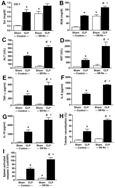
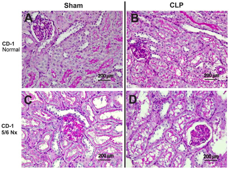
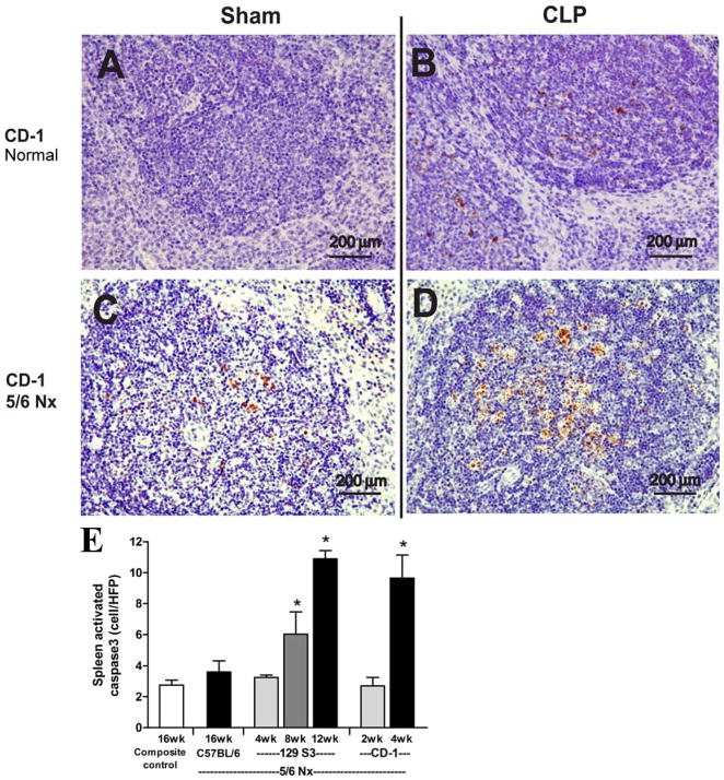
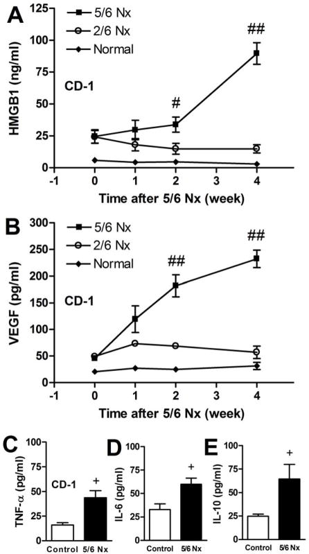
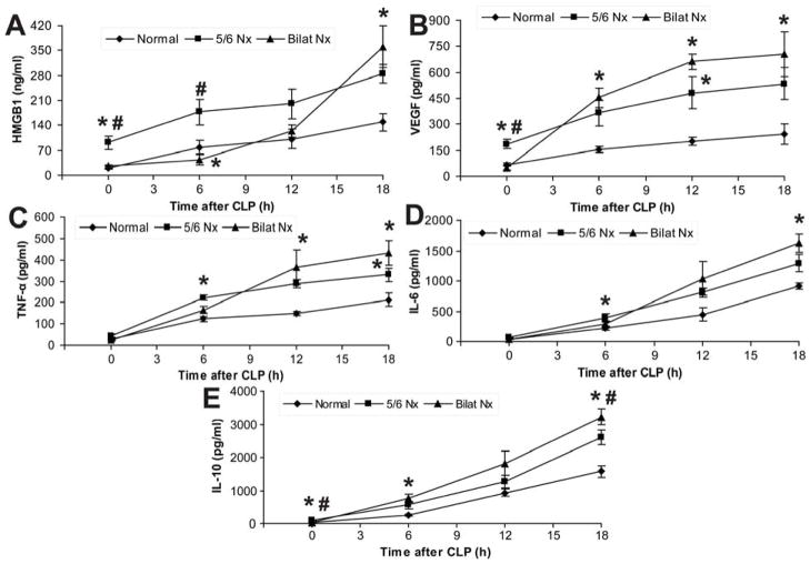
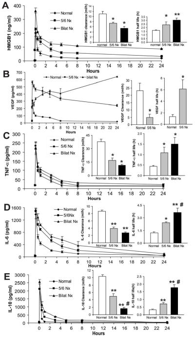
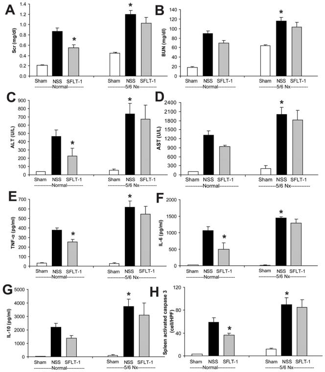
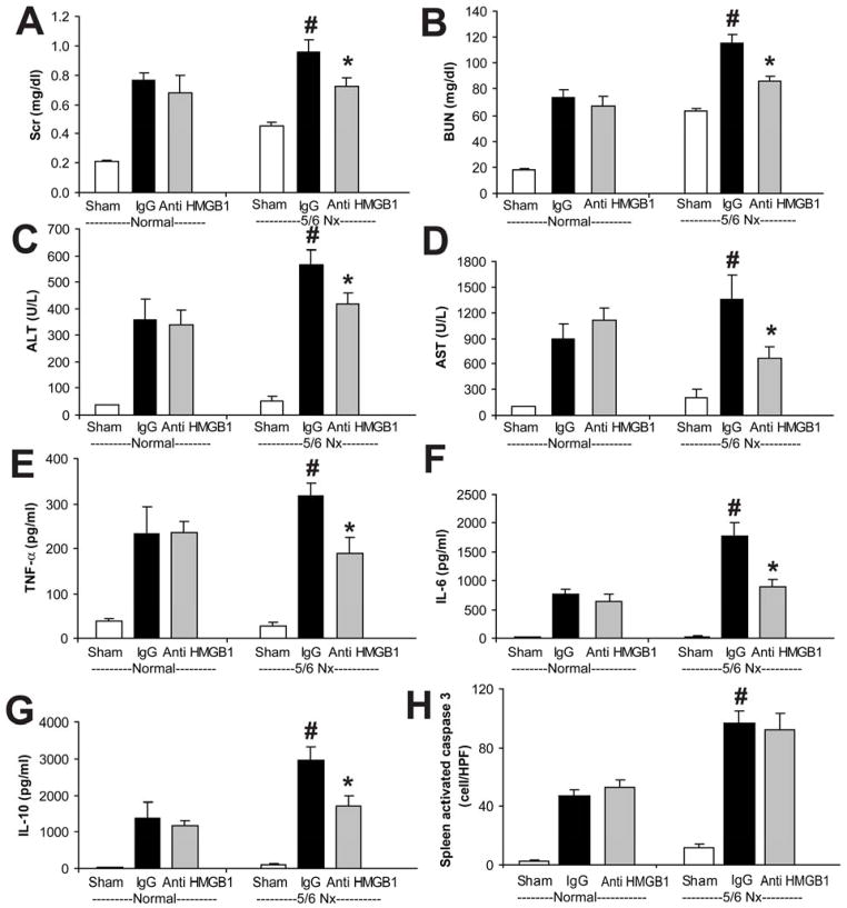
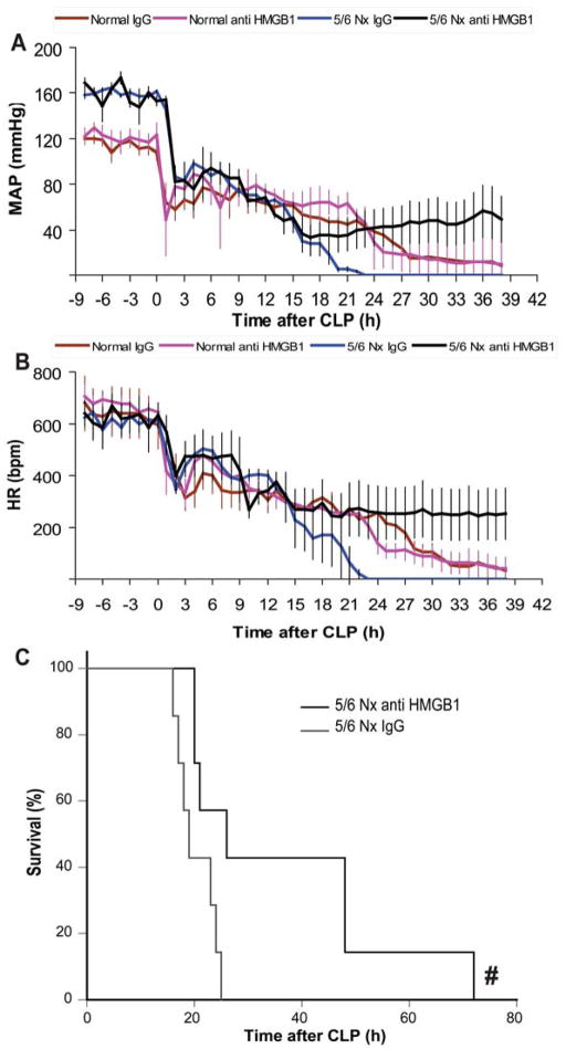
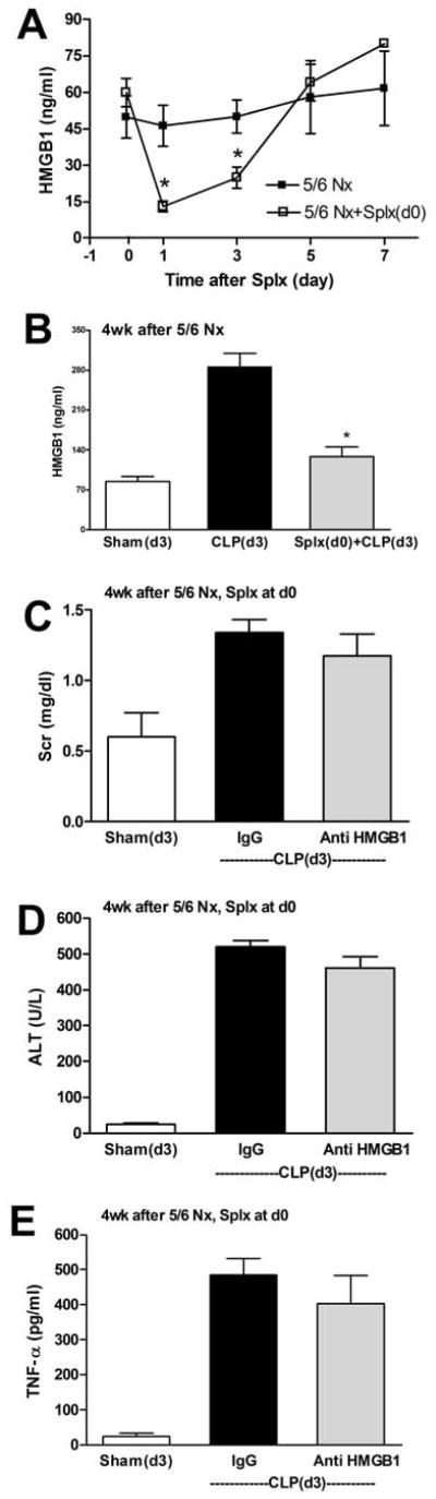
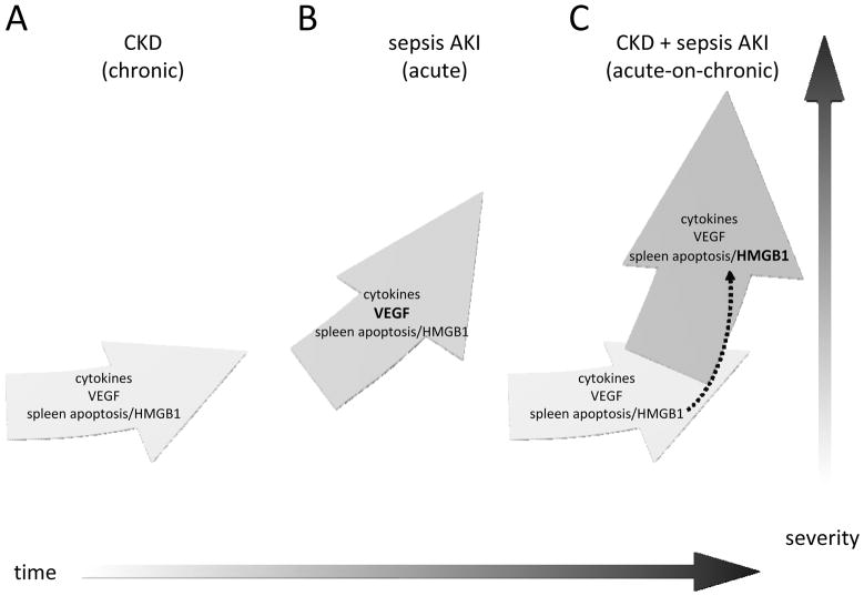
We have shown that folate-induced kidney dysfunction and interstitial fibrosis predisposes mice to sepsis mortality. Agents that increase survival in normal septic mice were ineffective in a two-stage kidney disease model. Here we used the 5/6 nephrectomy mouse model of progressive chronic kidney disease (CKD) to study how CKD affects acute kidney injury (AKI) induced by sepsis. We induced sepsis using cecal ligation and puncture and found that the presence of CKD intensified the severity of kidney and liver injury, cytokine release, and splenic apoptosis. Accumulation of High Mobility Group Box Protein-1 (HMGB1; a late proinflammatory cytokine released from apoptotic cells), vascular endothelial growth factor (VEGF), tumor necrosis factor (TNF)-α, interleukin (IL)-6, or IL-10 was increased in CKD or sepsis alone and to a greater extent in CKD-sepsis. Only part of the increase was explained by decreased renal clearance. Surprisingly, we found splenic apoptosis in CKD, even in the absence of sepsis. Although VEGF neutralization with soluble fms-like tyrosine kinase 1 (sFLT-1) (a soluble VEGF receptor) effectively treated sepsis, it was ineffective against CKD-sepsis. A single dose of HMGB1-neutralizing antiserum administered 6 h after sepsis alone was ineffective; however, CKD-sepsis was attenuated by anti-HMGB1. Splenectomy transiently decreased circulating HMGB1 levels, reversing the effectiveness of anti-HMGB1 treatment on CKD-sepsis. Thus, progressive CKD increases the severity of sepsis, in part, by reducing the renal clearance of several cytokines. CKD-induced splenic apoptosis and HMGB1 release could be important common mediators for both CKD and sepsis.


Figures

Figure 1
Figure 1. Widespread exacerbation of sepsis outcomes by CKD
CKD was induced by 5/6Nx in CD-1 mice, and CLP surgery was performed 4 weeks later. Organ injury was measured at 18 h after sham (white bar) or CLP (black bar) surgery. Renal function was determined by serum creatinine (A) and BUN (B), liver function was determined by ALT (C), AST (D), inflammation was determined by serum cytokine levels (TNF-α, IL-6, IL-10) (E–G), renal injury was determined by semi-quantitative measurement of renal vacuolized tubules (H), and splenic apoptosis (I) was measured by activated caspase 3 (n=6–7). *, P<0.05 vs. control sham; #, P<0.05 vs. control CLP; +, P<0.05 vs. 5/6 Nx sham.
Figure 2
Figure 2. CKD increases sepsis-induced renal tubular vacuolization
Representative images of periodic acid-Schiff-stained renal cortex in normal CD-1 mice (A, B), or CD-1 mice 4 weeks after 5/6 Nx (G, H) in sham (A, C) and CLP (B, D). Black bar corresponds to 200 μm.
Figure 3
Figure 3. CKD enhances sepsis-induced splenic apoptosis
CD-1 mice were untreated (normal, A, B) or subjected to 5/6 Nx (C, D) 4 weeks prior to sham (A, C) or CLP surgery (B, D). Representative images of spleen stained for activated caspase3 are shown (A–D); black bar corresponds to 200 μm. Number of activated caspase3 positive cells per high powered field in spleen of composite control at 16 weeks (n=9); C57BL/6 5/6 Nx at 16 weeks (n=4); 129S3 5/6 Nx at 4, 8, 12 weeks (n= 4/group); CD-1 5/6 Nx at 2, 4 weeks (n=4/group)
Figure 4
Figure 4. Accumulation of HMGB1, and VEGF, but not TNF-α, IL-6 and IL-10 during CKD (5/6 Nx) progression
Time course of serum HMGB1 (A); and VEGF (B) during the course of 5/6 Nx-induced CKD (n= 4–6/group). Repeated measures ANOVA with post-hoc (Tukey) comparisons were performed (## P<0.0001 vs. 2/6 Nx, #P<0.05 vs. 2/6 Nx). Serum levels of TNF-α (C), IL-6 (D), IL-10 (E) were measured in a composite of normal and 2/6 Nx controls vs. 5/6 Nx at 4 weeks (+ P<0.05 vs. control by one-way ANOVA).
Figure 5
Figure 5. Time course of serum cytokines (HMGB1, VEGF, TNF-α, IL-6 and IL-10) following CLP with or without pre-existing kidney impairment
Serum HMGB1 (A), VEGF (B), TNF-α (C), IL-6 (D) and IL-10 (E) at indicated time points in CD-1 mice after normal-CLP, 5/6 Nx-CLP or bilateral Nx-CLP. (n = 4–5/time point). By repeated measures ANOVA all except IL-6 had significant time x nephrectomy interactions; post-hoc (Tukey) comparisons: * P<0.05 vs. normal-CLP, # P<0.05 vs. 5/6 Nx-CLP.
Figure 6
Figure 6. Comparison of cytokine clearance/half-life in normal, 5/6 Nx and bilateral Nx mice
After injection of exogenous recombinant HMGB1 (A), VEGF (B), TNF-α (C), IL-6 (D) or IL-10 (E) serum cytokine concentrations were measured at different times for area under the curve (AUC), clearance, and half-life calculations. (n = 4–5/group) * P<0.05 vs. normal, ** P<0.03 vs. normal, # P <0.05 vs. 5/6 Nx.
Figure 7
Figure 7. sFLT-1 attenuated sepsis severity in normal-CLP but not in 5/6 Nx-CLP mice
CD-1 mice 4 weeks after 5/6 Nx or normal controls were subjected to sham surgery (white bars) or CLP, then injected at 0, 3, 6, and 9 h after CLP with saline (NSS, black bars) or soluble FLT-1 (sFLT-1, 33.3 mg/kg i.v., gray bars). The following were measured 18 h post-CLP: renal injury (Scr, BUN) (A, B), liver injury (ALT, AST) (C, D), inflammatory cytokines (TNF-α, IL-6, IL-10) (E–G), and splenic apoptosis (H) (n = 4–6/group). By ANOVA, there was no significant interaction between CKD-sepsis and treatment; post-hoc comparisons (Holm-Sidak) comparisons: * P<0.05 vs. normal-CLP + normal saline.
Figure 8
Figure 8. Anti-HMGB1 attenuated sepsis in 5/6 Nx but not normal mice
CD-1 mice 4 weeks after 5/6 Nx or normal controls were subjected to sham surgery (white bars) or CLP, then injected at 6 h after CLP with control rabbit IgG (IgG, black bars) or anti-HMGB1 (3.6 mg/kg, i.p., gray bars). The following were measured 18 h post-CLP: renal injury (Scr, BUN) (A, B), liver injury (ALT, AST) (C, D), inflammatory cytokines (TNF-α, IL-6, IL-10) (E–G), and splenic apoptosis (H) (n = 4–6/group). By ANOVA, there was significant interaction between CKD-sepsis and treatment for BUN, AST, TNFα, IL-6, and splenic apoptosis; post-hoc comparisons (Holm-Sidak) comparisons: * P<0.05 vs. normal-CLP + rabbit IgG, # P<0.05 vs. 5/6 Nx-CLP + rabbit IgG.
Figure 9
Figure 9. Anti-HMGB1 improved sepsis-induced hypotension, bradycardia and survival after 5/6 Nx and sepsis
Telemetric recording of conscious mean arterial pressure (MAP) (A) and heart rate (HR) (B) of normal (red, pink) or 5/6 Nx mice (blue, black) subjected to CLP, and after 6 hours injected with rabbit IgG control (red, blue) or anti-HMGB1 (pink, black) (n=4/group). By repeated measures ANOVA p<0.0001 for either MAP or HR; post-hoc (Tukey) comparisons 5/6 Nx-CLP + IgG control vs. 5/6 Nx-CLP + anti-HMGB1 was significant 20–38 h post-CLP (MAP) and 15–38 h, except 30,35, and 36 h (HR). Survival curve (C) of 5/6 Nx mice subjected to CLP, then treatment 6 h later with rabbit IgG control (gray) or anti-HMGB1 (black) (n = 7/group). # P<0.05 anti-HMGB1 vs. rabbit IgG control (Kaplan-Meier analysis)
Figure 10
Figure 10. Splenectomy transiently decreases HMGB1 and renders anti-HMGB1 ineffective in treating CKD-sepsis
CD-1 mice were subjected to 5/6 Nx, and after 4 weeks, splenectomy was performed at day 0. Serum HMGB1 (A) was measured in 5/6 Nx controls (black squares) or 5/6 Nx-splenectomy (white squares). Loss of CKD-sepsis-stimulated serum HMGB1 (B): four weeks after 5/6 Nx mice were subjected to splenectomy (gray bar), and after three days sham surgery (white bar) or CLP (black bar, gray bar) was performed, and serum HMGB1 was measured after 18 h. Outcomes of CKD-sepsis (see also Supplemental Fig 4): four weeks after 5/6 Nx mice were subjected to splenectomy (day 0), and after three days sham surgery (white bars) or CLP (black bars, gray bars) was performed, followed by administration of control IgG (black bars) or anti-HMGB1 (gray bars) 6 h later, then measurement of Scr (C), ALT (D), or TNFα (E) at 18 h.
Figure 11
Figure 11. Proposed framework for Acute-on-Chronic kidney disease
Chronic kidney disease progresses slowly (A), and after a septic insult the trajectory accelerates (C) relative to uncomplicated sepsis-AKI (B). This acceleration may be attributable, in part, to 1) to decreased GFR, which increases levels of cytokines, VEGF, and HMGB1, and 2) existing spleen apoptosis and/or HMGB1 during CKD may enhance the sepsis-induced increase in HMGB1 (dashed arrow). A mechanistic shift occurs when CKD and sepsis-AKI are combined; Acute-on-Chronic kidney disease is distinct from the sum of its parts. Anti-VEGF treatment, which is effective for sepsis-AKI, is no longer effective in Acute-on-Chronic kidney disease; and anti-HMGB1 treatment, which was ineffective for uncomplicated sepsis-AKI, is effective for Acute-on-Chronic kidney disease.

References

    1. Martin GS, Mannino DM, Eaton S, Moss M. The epidemiology of sepsis in the United States from 1979 through 2000. N Engl J Med. 2003 Apr 17;348(16):1546–54. - PubMed
    1. Doi K, Leelahavanichkul A, Yuen PS, Star RA. Animal models of sepsis and sepsis-induced kidney injury. J Clin Invest. 2009 Oct;119(10):2868–78. - PMC - PubMed
    1. Dyson A, Singer M. Animal models of sepsis: why does preclinical efficacy fail to translate to the clinical setting? Crit Care Med. 2009 Jan;37(1 Suppl):S30–7. - PubMed
    1. Esmon CT. Why do animal models (sometimes) fail to mimic human sepsis? Crit Care Med. 2004 May;32(5 Suppl):S219–22. - PubMed
    1. Riedemann NC, Guo RF, Ward PA. The enigma of sepsis. J Clin Invest. 2003 Aug;112(4):460–7. - PMC - PubMed

Publication types