. 2010 Oct 18:1:169.
doi: 10.3389/fpsyg.2010.00169. eCollection 2010.

Neural markers of opposite-sex bias in face processing

Affiliations

Neural markers of opposite-sex bias in face processing

Alice Mado Proverbio et al. Front Psychol. .

Abstract

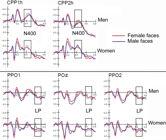
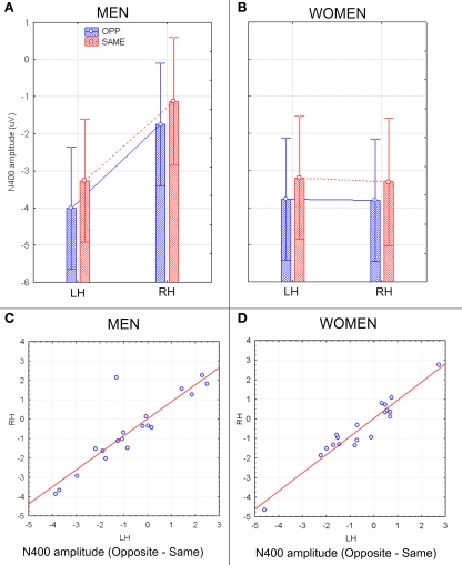
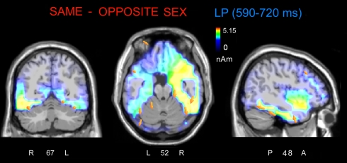
Some behavioral and neuroimaging studies suggest that adults prefer to view attractive faces of the opposite sex more than attractive faces of the same sex. However, unlike the other-race face effect (Caldara et al., 2004), little is known regarding the existence of an opposite-/same-sex bias in face processing. In this study, the faces of 130 attractive male and female adults were foveally presented to 40 heterosexual university students (20 men and 20 women) who were engaged in a secondary perceptual task (landscape detection). The automatic processing of face gender was investigated by recording ERPs from 128 scalp sites. Neural markers of opposite- vs. same-sex bias in face processing included larger and earlier centro-parietal N400s in response to faces of the opposite sex and a larger late positivity (LP) to same-sex faces. Analysis of intra-cortical neural generators (swLORETA) showed that facial processing-related (FG, BA37, BA20/21) and emotion-related brain areas (the right parahippocampal gyrus, BA35; uncus, BA36/38; and the cingulate gyrus, BA24) had higher activations in response to opposite- than same-sex faces. The results of this analysis, along with data obtained from ERP recordings, support the hypothesis that both genders process opposite-sex faces differently than same-sex faces. The data also suggest a hemispheric asymmetry in the processing of opposite-/same-sex faces, with the right hemisphere involved in processing same-sex faces and the left hemisphere involved in processing faces of the opposite sex. The data support previous literature suggesting a right lateralization for the representation of self-image and body awareness.

Keywords: ERPs; body awareness; face coding; hemispheric asymmetry; self-representation; sex differences; social cognition; visual perception.


Figures

Figure 1
Figure 1
Timeline of stimulus presentation. Each picture was displayed for 800 ms and followed by a random ISI. The task consisted of responding to landscapes as quickly and accurately as possible.
Figure 2
Figure 2
ERP waveforms recorded at left, right and midline centro–parietal and occipito–parietal sites in women (N = 19) and men (N = 19) in response to faces of female and male individuals. Both genders exhibited an enlarged N400 to opposite-sex faces and a large late positivity (LP) to same-sex faces.
Figure 3
Figure 3
Grand-average ERP waveforms (N = 38) recorded at centro–parietal and occipito–parietal sites (corresponding to the sites included in the N400 and LP ANOVAs) as a function of the face gender (opposite vs. same), along with the difference wave obtained by subtracting ERPs to same-sex faces from ERPs to opposite-sex faces.
Figure 4
Figure 4
(A,B) N400 peak latency values (in ms) with within-subjects standard errors recorded in men (A) and women (B) as a function of face gender and cerebral hemisphere. No effect of sex of viewers was found, but a significant opposite-sex bias was observed. (C,D) The N400 latency difference between opposite and same for men and women recorded at left and right centro–parietal sites. Negative values indicate expedite opposite-sex processing. Results were consistent across subjects and sex groups.
Figure 5
Figure 5
N400 peak amplitude values (in μV) with within-subjects standard errors recorded in men (A) and women (B) as a function of face gender and cerebral hemisphere. A strong hemispheric asymmetry is visible in men (A) but not women (B). (C,D) Scatter plots displaying individual values of N400 amplitudes (in μV) obtained by subtracting opposite–same responses recorded in men (C) and women (D) at left and right centro/parietal sites. Negative values indicate an enhanced brain processing of opposite-sex faces.
Figure 6
Figure 6
(A) Isocontour voltage topographical maps (top, front, and side views) of the face/gender effect obtained by subtracting ERPs associated with same-sex faces from ERPs associated with opposite-sex faces during the 400–450 ms time window (N400). (B) Topographical maps obtained by subtracting ERPs associated with opposite-sex faces from ERPs associated with same-sex faces during the 590–720 ms time window (LP).
Figure 7
Figure 7
The swLORETA inverse solution applied on the opposite-/same-sex difference wave during the time window 400–500 ms, which corresponds to the peak of the N400 response. The electromagnetic dipoles are shown as yellow arrows and indicate the position, orientation, and magnitude of the dipole-modeling solution applied to the ERP difference wave in this specific time window.
Figure 8
Figure 8
(A,B) LP mean-amplitude values (in μV) with within-subjects standard errors recorded in men (A) and women (B) as a function of face gender and recording site. (C,D) Scatter plot displaying individual values of LP component obtained by subtracting same–opposite responses recorded in men (C) and women (D) at left and right occipito–parietal sites. Positive values indicate larger cerebral responses to same-sex faces.
Figure 9
Figure 9
The swLORETA inverse solution applied on the same/opposite-sex difference wave during the time window 590–720 ms, which corresponds to the peak of the late positivity (LP). The electromagnetic dipoles are shown as yellow arrows and indicate the position, orientation, and magnitude of the dipole-modeling solution applied to the ERP difference wave in this specific time window.

Similar articles

Cited by

References

    1. Balas B., Nelson C. A. (2010). The role of face shape and pigmentation in other-race face perception: an electrophysiological study. Neuropsychologia 48, 498–50610.1016/j.neuropsychologia.2009.10.007 - DOI - PMC - PubMed
    1. Brown C., Hagoort P. (1993). The processing nature of the N400: evidence from masked priming. J. Cogn. Neurosci. 5, 34–4410.1162/jocn.1993.5.1.34 - DOI - PubMed
    1. Byatt G., Rhodes G. (2004). Identification of own-race and other-race faces: implications for the representation of race in face space. Psychon. Bull. Rev. 11, 735–741 - PubMed
    1. Caldara R., Rossion B., Bovet P., Hauert C. A. (2004). Event-related potentials and time course of the “other-race” face classification advantage. Neuroreport 9, 905–91010.1097/00001756-200404090-00034 - DOI - PubMed
    1. Conway C., Jones B. C., DeBruine L. M., Little A. C. (2008). Evidence for adaptive design in human gaze preference. Proc. Biol. Sci. 7, 63–6910.1098/rspb.2007.1073 - DOI - PMC - PubMed

LinkOut - more resources