

Lipopolysaccharide-Deficient Brucella Variants Arise Spontaneously during Infection
- PMID: 21833310
- PMCID: PMC3153030
- DOI: 10.3389/fmicb.2011.00054
Lipopolysaccharide-Deficient Brucella Variants Arise Spontaneously during Infection
Abstract
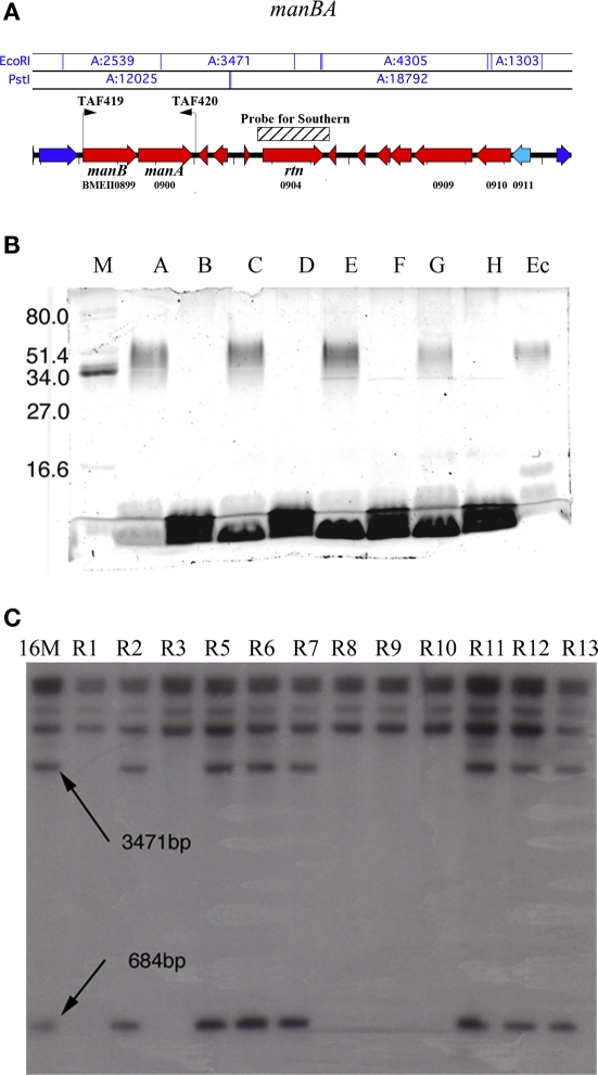
Lipopolysaccharide-deficient mutants of smooth Brucella species (rough mutants) have been shown to arise spontaneously in culture. However, in situ analysis of Brucella infected macrophages using antibody directed against O-polysaccharide suggested a loss of reactivity of Brucella consistent with the appearance of rough organisms, and a potential contribution to infection. The experiments reported describe the direct recovery of Brucella from macrophages infected in vitro and from the spleens of infected mice at a frequency similar to that described in vitro, suggesting that Brucella dissociation is not simply an in vitro artifact. The frequency of appearance of spontaneous rough organisms deficient in O-polysaccharide expression measured in vitro is approximately 2-3 logs higher than the appearance of mutation to antibiotic resistance, purine auxotrophy, or reversion of erythritol sensitive ΔeryC mutants to tolerance. Genetic trans-complementation using a plasmid-based expression of Brucella manBA successfully restored O-polysaccharide expression in only one-third of O-polysaccharide deficient spontaneous mutants. Suggesting that the appearance of rough mutants is caused by mutation at more than one locus. In addition, Sanger sequencing of the manBA structural genes detected multiple sequence changes that may explain the observed phenotypic differences. The presence of O-polysaccharide resulted in macrophage and neutrophil infiltration into the peritoneal cavity and systemic distribution of the organism. In contrast, rough organisms are controlled by resident macrophages or by extracellular killing mechanisms and rapidly cleared from this compartment consistent with the inability to cause disease. Loss of O-polysaccharide expression appears to be stochastic giving rise to organisms with biological properties distinct from the parental smooth organism during the course of infection.
Keywords: Brucella; O-polysaccharide; lipopolysaccharide; rough mutant.
Figures
Similar articles
-
Brucellosis vaccines: assessment of Brucella melitensis lipopolysaccharide rough mutants defective in core and O-polysaccharide synthesis and export.PLoS One. 2008 Jul 23;3(7):e2760. doi: 10.1371/journal.pone.0002760. PLoS One. 2008. PMID: 18648644 Free PMC article.
-
Spontaneous excision of the O-polysaccharide wbkA glycosyltranferase gene is a cause of dissociation of smooth to rough Brucella colonies.J Bacteriol. 2012 Apr;194(8):1860-7. doi: 10.1128/JB.06561-11. Epub 2012 Feb 10. J Bacteriol. 2012. PMID: 22328663 Free PMC article.
-
Brucella abortus rough mutants induce macrophage oncosis that requires bacterial protein synthesis and direct interaction with the macrophage.Infect Immun. 2006 May;74(5):2667-75. doi: 10.1128/IAI.74.5.2667-2675.2006. Infect Immun. 2006. PMID: 16622203 Free PMC article.
-
Rough vaccines in animal brucellosis: structural and genetic basis and present status.Vet Res. 2004 Jan-Feb;35(1):1-38. doi: 10.1051/vetres:2003037. Vet Res. 2004. PMID: 15099501 Review.
-
When the Going Gets Rough: The Significance of Brucella Lipopolysaccharide Phenotype in Host-Pathogen Interactions.Front Microbiol. 2021 Jul 15;12:713157. doi: 10.3389/fmicb.2021.713157. eCollection 2021. Front Microbiol. 2021. PMID: 34335551 Free PMC article. Review.
Cited by
-
Biological Characterization and DIVA Potential of Three Rough Brucella melitensis Vaccine Strains.Vaccines (Basel). 2025 Aug 13;13(8):857. doi: 10.3390/vaccines13080857. Vaccines (Basel). 2025. PMID: 40872942 Free PMC article.
-
Mechanism of Asp24 upregulation in Brucella abortus rough mutant with a disrupted O-antigen export system and effect of Asp24 in bacterial intracellular survival.Infect Immun. 2014 Jul;82(7):2840-50. doi: 10.1128/IAI.01765-14. Epub 2014 Apr 21. Infect Immun. 2014. PMID: 24752516 Free PMC article.
-
What have we learned from brucellosis in the mouse model?Vet Res. 2012 Apr 13;43(1):29. doi: 10.1186/1297-9716-43-29. Vet Res. 2012. PMID: 22500859 Free PMC article. Review.
-
Brucella abortus Strain 2308 Wisconsin Genome: Importance of the Definition of Reference Strains.Front Microbiol. 2016 Sep 29;7:1557. doi: 10.3389/fmicb.2016.01557. eCollection 2016. Front Microbiol. 2016. PMID: 27746773 Free PMC article.
-
Smooth to Rough Dissociation in Brucella: The Missing Link to Virulence.Front Cell Infect Microbiol. 2016 Jan 5;5:98. doi: 10.3389/fcimb.2015.00098. eCollection 2015. Front Cell Infect Microbiol. 2016. PMID: 26779449 Free PMC article.
References
-
- Alton G. G., Jones L. M., Pietz D. E. (1975). Laboratory techniques in brucellosis. Monogr. Ser. World Health Organ. 1–163 - PubMed
-
- Anderson T. D., Cheville N. F., Meador V. P. (1986a). Pathogenesis of placentitis in the goat inoculated with Brucella abortus. II. Ultrastructural studies. Vet. Pathol. 23, 227–239 - PubMed
-
- Anderson T. D., Meador V. P., Cheville N. F. (1986b). Pathogenesis of placentitis in the goat inoculated with Brucella abortus. I. Gross and histologic lesions. Vet. Pathol. 23, 219–226 - PubMed
-
- Barquero-Calvo E., Chaves-Olarte E., Weiss D. S., Guzman-Verri C., Chacon-Diaz C., Rucavado A., Moriyon I., Moreno E. (2007). Brucella abortus uses a stealthy strategy to avoid activation of the innate immune system during the onset of infection. PLoS ONE 2, e631.10.1371/journal.pone.0000631 - DOI - PMC - PubMed
LinkOut - more resources
Full Text Sources