. 2011 Aug 1;2(8):2372-82.
doi: 10.1364/BOE.2.002372. Epub 2011 Jul 25.

Differentiation of pancreatic cysts with optical coherence tomography (OCT) imaging: an ex vivo pilot study

Differentiation of pancreatic cysts with optical coherence tomography (OCT) imaging: an ex vivo pilot study

Nicusor Iftimia et al. Biomed Opt Express. .

Abstract

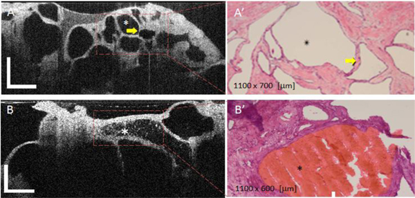
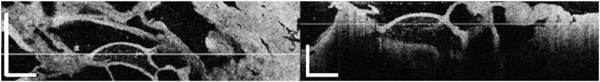
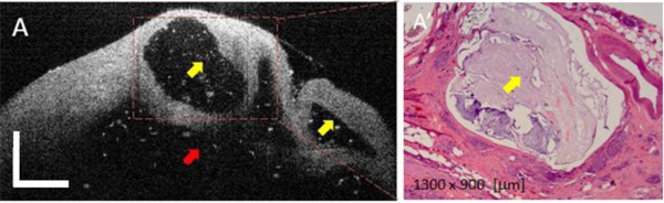
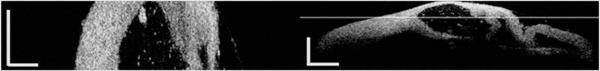
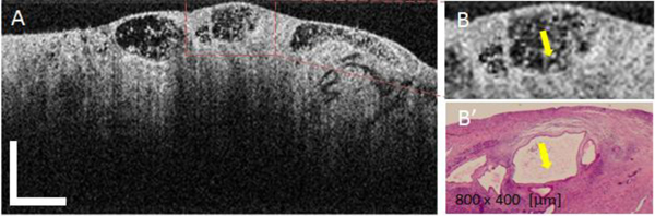
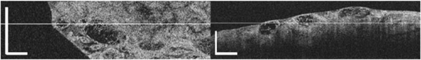
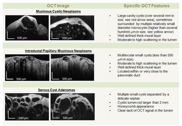
We demonstrate for the first time that optical coherence tomography (OCT) imaging can reliably distinguish between morphologic features of low risk pancreatic cysts (i.e., pseudocysts and serous cystadenomas) and high risk pancreatic cysts (i.e., mucinous cystic neoplasms and intraductal papillary mucinous neoplasms). In our study fresh pancreatectomy specimens (66) from patients with cystic lesions undergoing surgery were acquired and examined with OCT. A training set of 20 pathology-OCT correlated tissue specimens were used to develop criteria for differentiating between low and high risk cystic lesions. A separate (validation) set of 46 specimens were used to test the OCT criteria by three clinicians, blinded to histopathology findings. Histology was finally used as a 'gold' standard for testing OCT findings. OCT was able to reveal specific morphologic features of pancreatic cysts and thus to differentiate between low-risk and high-risk cysts with over 95% sensitivity and specificity. This pilot study suggests that OCT could be used by clinicians in the future to more reliably differentiate between benign and potentially malignant pancreatic cysts. However, in vivo use of OCT requires a probe that has to fit the bore of the pancreas biopsy needle. Therefore, we have developed such probes and planned to start an in vivo pilot study within the very near future.

Keywords: (170.0170) Medical optics and biotechnology; (170.4500) Optical coherence tomography; (170.4580) Optical diagnostics for medicine; (170.6935) Tissue characterization.


Figures

Fig. 1
Fig. 1
Simplified schematic (A) and photograph of the OCT system (B).
Fig. 2
Fig. 2
OCT appearance of SCAs (A) and (B) and corresponding histology (A', B'). The microcystic SCAs show multiple tiny cysts with well-defined outlines. The thin septae between the cysts (see arrow), creating a honeycomb appearance, shows homogenously high scattering. The cyst content usually lacks a scattering effect. Focal intraluminal scattering can be noted on some cysts (see asterisk in Fig. 2(B)) and it usually corresponds to a focus of intraluminal hemorrhage, as confirmed by histology (see red area Fig. 2(B′)). OCT scale bar = 500 μm.
Fig. 3
Fig. 3
(Media 1). Fly-through video of a SCA. (A) Enface view at a depth indicated by the line from the cross-sectional image. (B) cross-sectional view. The raw video was acquired at 20 fps. Scale bar = 500 μm.
Fig. 4
Fig. 4
OCT appearance of a mucinous cyst (A) and corresponding histology (B). Yellow arrows indicate the presence of smaller size (daughter) cysts, while the red arrow indicates the main cystic cavity. The cystic content (mucin) shows some scattering due the presence of dead epithelial cells. OCT scale bar = 500 μm.
Fig. 5
Fig. 5
(Media 2). Fly-through video of a MCN. (A) Enface view at a depth indicated by the line from the cross-sectional view. (B) Cross-sectional view. The raw video was acquired at 20 fps. Scale bar = 500 μm.
Fig. 6
Fig. 6
OCT appearance of mucinous IPMN cysts (A), magnified ROI (B), and corresponding histology (B'). Yellow arrows indicate the areas of highly scattering mucin within the cyst fluid. OCT Scale bar = 500 μm.
Fig. 7
Fig. 7
(Media 3). Fly-through video of a IPMN. (A) Enface view. (B) Cross-sectional view. The raw video was acquired at 20 fps. Scale bar = 500 μm.
Fig. 8
Fig. 8
OCT diagnostic criteria for pancreatic cysts.

References

    1. Brugge W. R., Lauwers G. Y., Sahani D., Fernandez-del Castillo C., Warshaw A. L., “Cystic neoplasms of the pancreas,” N. Engl. J. Med. 351(12), 1218–1226 (2004).10.1056/NEJMra031623 - DOI - PubMed
    1. Lee K. S., Sekhar A., Rofsky N. M., Pedrosa I., “Prevalence of incidental pancreatic cysts in the adult population on MR imaging,” Am. J. Gastroenterol. 105(9), 2079–2084 (2010).10.1038/ajg.2010.122 - DOI - PubMed
    1. Sahani D. V., Miller J. C., Fernàndez del Castillo C., Brugge W. R., Thrall J. H., Lee S. I., “Cystic pancreatic lesions: classification and management,” J. Am. Coll. Radiol. 6(5), 376–380 (2009).10.1016/j.jacr.2008.10.004 - DOI - PubMed
    1. Wargo J. A., Fernandez-del-Castillo C., Warshaw A. L., “Management of pancreatic serous cystadenomas,” Adv. Surg. 43(1), 23–34 (2009).10.1016/j.yasu.2009.03.001 - DOI - PubMed
    1. Federle M. P., McGrath K. M., “Cystic neoplasms of the pancreas,” Gastroenterol. Clin. North Am. 36(2), 365–376, ix (2007).10.1016/j.gtc.2007.03.014 - DOI - PubMed

LinkOut - more resources