. 2011 Oct;136(4):399-411.
doi: 10.1007/s00418-011-0852-9. Epub 2011 Aug 11.

Attachment of HeLa cells during early G1 phase

Affiliations

Attachment of HeLa cells during early G1 phase

Maarten J A Moes et al. Histochem Cell Biol. 2011 Oct.

Abstract

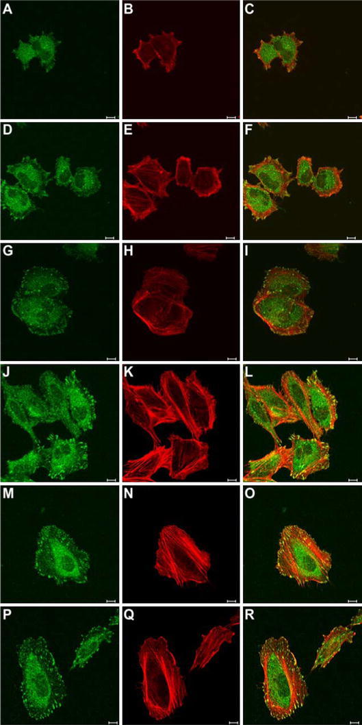
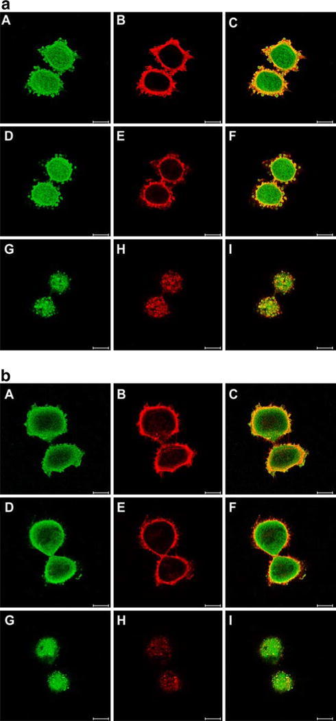
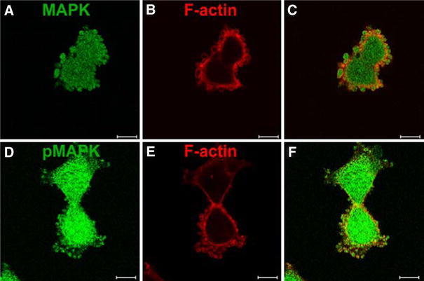
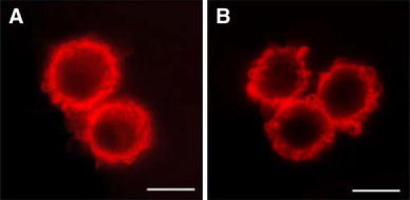
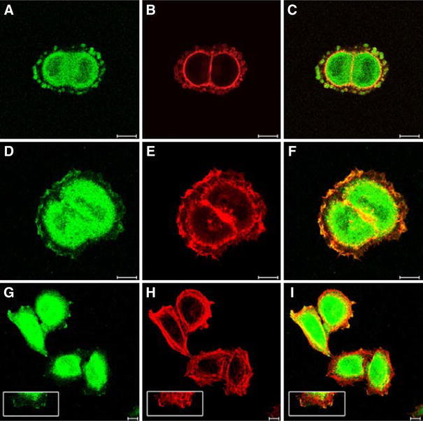
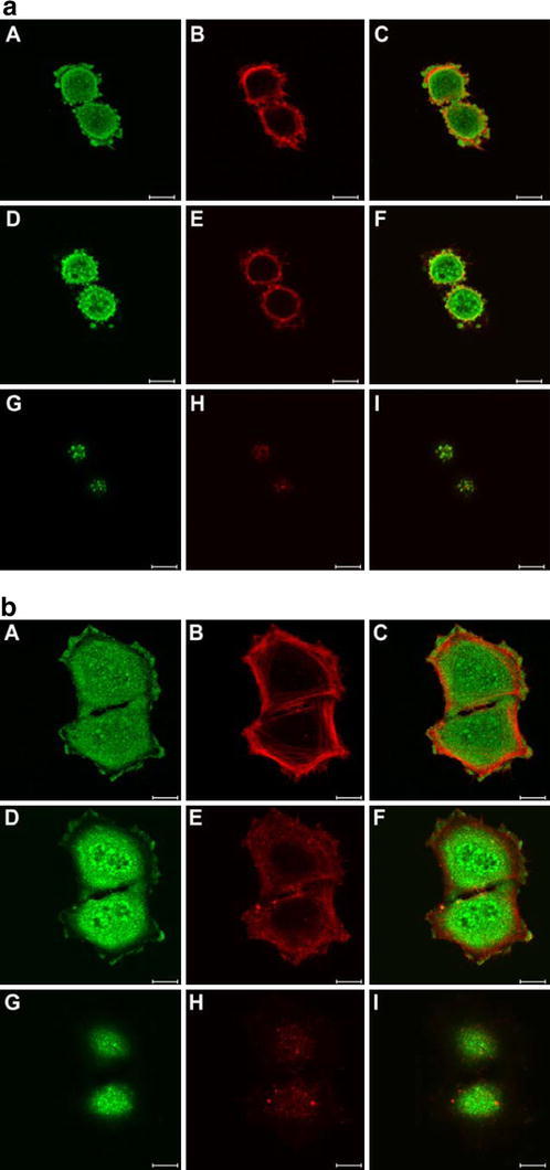
Both growth factor directed and integrin dependent signal transduction were shown to take place directly after completion of mitosis. The local activation of these signal transduction cascades was investigated in early G1 cells. Interestingly, various key signal transduction proteins were found in blebs at the cell membrane within 30 min after mitosis. These membrane blebs appeared in round, mitotic-like cells and disappeared rapidly during spreading of the cells in G1 phase. In addition to tyrosine-phosphorylated proteins, the blebs contained also phosphorylated FAK and phosphorylated MAP kinase. The formation of membrane blebs in round, mitotic cells before cell spreading is not specific for mitotic cells, because similar features were observed in trypsinized cells. Just before cell spreading also these cells exhibited membrane blebs containing active signal transduction proteins. Inhibition of signal transduction did not affect membrane bleb formation, suggesting that the membrane blebs were formed independent of signal transduction.


Figures

Fig. 1
Fig. 1
Localization of tyrosine phosphorylated proteins and F-actin in early G1 phase HeLa cells. HeLa cells were chemically fixed 30 min after mitotic shake off and stained for PY100 (green) and F-actin (red) as described in “Materials and methods”. The upper panel (ac) represents the initial stage after mitosis. Cells exhibit blebs of the cell membrane that contain F-actin and are enriched with proteins that are phosphorylated at tyrosine residues. Cells that are spread further exhibit small focal adhesions and thin actin stress fibers (d–f). Cells that show small focal adhesions contain no blebs of the cell membrane. Bar represents 10 μm
Fig. 2
Fig. 2
Localization of tyrosine phosphorylated proteins and F-actin in G1 phase of HeLa cells. HeLa cells were synchronized by mitotic shake-off and replated for 1, 2, 3, 4, 5 and 6 h, respectively, as indicated. Labelling of tyrosine phosphorylated proteins [PY100 labelling (green)] is localized throughout the cytoplasm and in focal adhesions. F-actin [Phalloidin labelling (red)] is present in focal adhesions and stress fibers. Focal adhesions increase both in size and in number (a, d, g, j, m, p). During time, the actin stress fibers become more apparent (b, e, h, k, n, q). Bar represents 10 μm
Fig. 3
Fig. 3
Localization of phosphorylated FAK during early G1 phase in HeLa cells. a HeLa cells were synchronized by mitotic shake-off and replated for 40 min followed by chemical fixation as described under “Materials and methods”. Phospho-FAK397 (green) and F-actin (red) were labeled as described under “Materials and methods”. Three optical sections of the same cells are represented: A basal side, B middle part and C apical side. P-FAK397 was detected in membrane blebs at the cell surface cells. The blebs contained F-actin, and P-FAK397 localized in membrane blebs was surrounded with a coat of F-actin (a C, F). a D, E, F represents an optical section through the center of the cell that clearly exhibits blebs at the edge of the cell. The blebs of the membrane were present on the whole cell surface, from the basal side (a A, B, C) to the apical side (a G, H, I). Bar represents 10 μm. b Synchronized HeLa cells that were chemically fixed 1 h after replating were stained for Phospho-FAK397 (green) and F-actin (red). Three optical sections of the same cells are represented: A basal side, B middle part and C apical. After 60 min, the number of blebs decreased at the apical side of cells (b G, H, I vs. a G, H, I). Optical sections through the center of the cell revealed no blebs of the membrane (b D, E, F) in contrast to sections 40 min after mitosis (a D, E, F). However, in the cells that were fixed after 60 min, some blebs were observed that contain P-FAK397 at the basal side of cells (b A, B, C). Bar represents 10 μm
Fig. 4
Fig. 4
Localization of MAP kinase and phosphorylated MAP kinase during early G1 phase in HeLa cells. HeLa cells were synchronized, replated and chemically fixed as described under “Material and methods” and Legends of Fig. 3. Subsequently cells were labelled for MAPK or phospho-MAPK labelling (green) and F-actin labeling (red). MAPkinase (a) and phospho-MAPkinase (d) are localized in blebs of the cell membrane that can be distinguished from the rest of the cell by the packaging in F-actin (b, e). Bar represents 10 μm
Fig. 5
Fig. 5
Blebs of the cell membrane in post-mitotic cells in a random growing cell cultures of CHO (a) and HeLa cells (b). Blebs of the cell membrane were detected by labelling for F-actin. Bar represents 10 μm
Fig. 6
Fig. 6
Spreading of trypsinized HeLa and CHO cells. a Cells were trypsinized according general procedures and allowed to spread for 30 or 60 min after replating. Subsequently cells were chemically fixed and labelled for PY100 (green) and F-actin (red) as described under “Materials and methods”. Bar represents 10 μm. b Effect of cell substratum on cell spreading and membrane bleb formation in CHO cells. CHO cells were trypsinized and plated on fibronectin and poly-l-lysine, respectively, in the absence or presence of serum for 10 or 20 min as indicated. Subsequently the fraction of the initial plated cells was calculated that had a spread or round morphology and the fraction of initial plated cells that contained membrane blebs. Because the cells were allowed to attach for only 10 or 20 min, non-attached cells are not included in the calculation
Fig. 7
Fig. 7
Localization of cPLA2α in G1 phase HeLa cells. HeLa cells were synchronized, replated and chemically fixed as described under “Materials and methods” and Legends of Fig. 3. Subsequently cells were labelled for cPLA2α (green) and F-actin (red). Chemical fixation was performed 30 min (ac), 1 h (d–f), and 4 h (g–i) after replating of mitotic cells. Bar represents 10 μm
Fig. 8
Fig. 8
Localization of phospho-cPLA2α in G1 phase HeLa cells. HeLa cells were synchronized, replated and chemically fixed as described under “Materials and methods” and Legends of Fig. 3. Subsequently cells were labelled for phospho-cPLA2α (green) and F-actin (red). a 30 min after replating, blebs of the cell membrane containing labelling for phospho-cPLA2α are localized at the basal side (A, B, C) as well as at the apical side of cells (G, H, I). b 2 h after replating, cells exhibit no blebs of the cell membrane at the apical side of cells (G, H, I) and blebs have increased in size or fused together at the basal side of cells (A, B, C). Bar represents 10 μm
Fig. 9
Fig. 9
Effect of inhibition of the MAPkinase pathway on membrane bleb formation. HeLa cells were synchronized, replated for 30 or 60 min as indicated and chemically fixed as described under “Materials and methods”. Subsequently cells were labelled for F-actin (red) as described above. The MEK1 and MEK2 inhibitor pd98059 was added immediately after mitotic shake-off. In the presence of MEK1 and MEK2 inhibitor pd98059 (b, d), blebs of the membrane are present comparable to non-treated cells (a, c). Bar represents 10 μm

References

    1. Badley RA, Woods A, Carruthers L, Rees DA. Cytoskeleton changes in fibroblast adhesion and detachment. J Cell Sci. 1980;43:379–390. - PubMed
    1. Bohmer RM, Scharf E, Assoian RK. Cytoskeletal integrity is required throughout the mitogen stimulation phase of the cell cycle and mediates the anchorage-dependent expression of cyclin D1. Mol Biol Cell. 1996;7:101–111. - PMC - PubMed
    1. Boonstra J, Moes MJ. Signal transduction and actin in the regulation of G1-phase progression. Crit Rev Eukaryot Gene Expr. 2005;15:255–276. - PubMed
    1. Boonstra J, Mummery CL, Tertoolen LJG, van der Saag PT, de Laat SW. Cation transport and growth regulation in neuroblastoma cells. Modulations of K+ transport and electrical properties during the cell cycle. J Cell Physiol. 1981;107:75–83. doi: 10.1002/jcp.1041070110. - DOI - PubMed
    1. Bottazzi MA, Buzzai M, Zhu X, Desdouets C, Brechot C, Assoian RK. Distinct effects of mitogens and the actin cytoskeleton on CREB and pocket protein phosphorylation control the extent and timing of cyclin A promoter activity. Mol Cell Biol. 2001;21:7607–7616. doi: 10.1128/MCB.21.22.7607-7616.2001. - DOI - PMC - PubMed

Publication types

Substances

LinkOut - more resources